Supplement Review #5: Theacrine


Background

Caffeine is the most commonly used nootropic and cognitive-enhancing drug in the world. It is found in nearly every energy drink, pre-workout, tea, and coffee. It is known for antagonizing the adenosine receptor, thus allowing the user to remain alert. However, it also has many other health benefits. Besides the benefits of caffeine, the biggest problem with chronic administration is tolerance buildup. A daily dose of 50-100mg taken for a few weeks no longer elicits the same response, so a new dose of 150-200mg is needed. This tolerance builds up extremely fast, to the point that many people are overconsuming caffeine and turning it into a harmful compound. The truth is, caffeine is wonderful and, when taken correctly, can have tremendous benefits for health and productivity. Fortunately, there are other compounds like caffeine that allow for a caffeine “break” or enhancement.

Caffeine is the most commonly used nootropic and cognitive-enhancing drug in the world. It is found in nearly every energy drink, pre-workout, tea, and coffee. It is known for antagonizing the adenosine receptor, thus allowing the user to remain alert. However, it also has many other health benefits. Besides the benefits of caffeine, the biggest problem with chronic administration is tolerance buildup. A daily dose of 50-100mg taken for a few weeks no longer elicits the same response, so a new dose of 150-200mg is needed. This tolerance builds up extremely fast, to the point that many people are overconsuming caffeine and turning it into a harmful compound. The truth is, caffeine is wonderful and, when taken correctly, can have tremendous benefits for health and productivity. Fortunately, there are other compounds similar to caffeine that allow for a caffeine “break” or enhancement when taken in combination.

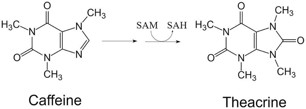
One such compound is theacrine, a naturally occurring purine alkaloid found in Camellia Kucha, a common tea whose leaves also produce caffeine at an earlier time point. Caffeine is converted to theacrine via S-adenosyl-l-methionine (SAM) in the leaves of Camellia Kucha. Theacrine acts like caffeine but has much more potential for chronic administration, meaning long-term administration of theacrine does not result in tolerance.

(Sheng et al., 2020)

Mechanisms of Action

Theacrine acts through many pathways in the body, although its primary target in eliciting a wakefulness state is the adenosine receptor. Theacrine acts as an adenosine receptor antagonist or blocker, which blocks adenosine from binding and illiciting the signaling cascade that follows. When adenosine builds up, it acts to bind (agonize) the adenosine receptors, which promote drowsiness. By blocking the adenosine receptor, theacrine blocks the ability for adenosine to bind, thus preventing adenosine-induced drowsiness. Theacrine also seems to regulate blood glucose metabolism and restore 5-HTP, a vital molecule in serotonergic signaling. Interestingly, theacrine also restores dopamine levels in certain regions of the brain.

(Sheng et al., 2020)

Benefits

Theacrine is a popular compound known as an alternative to caffeine. Although their mechanisms of action are somewhat similar, it is important to note that theacrine does not seem to increase central nervous system excitation to the extent that caffeine does. In terms of half-life, the major advantage of theacrine over caffeine and other stimulants is the long 20-hour half-life. Compared with caffeine’s 5-hour half-life, a one-time dose of theacrine will be sufficient for the entire day.

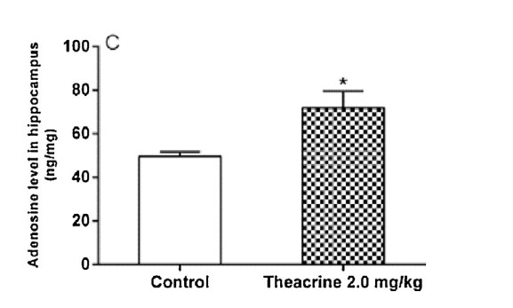
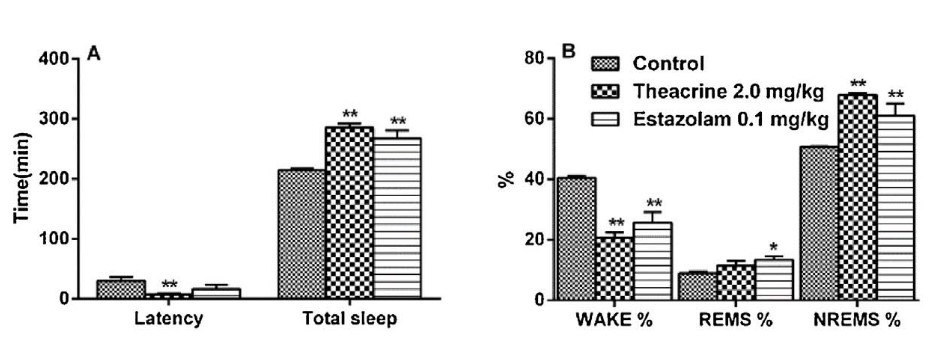
The primary effect that comes from theacrine administration is wakefulness. This is due to the blocking of the adenosine receptor by theacrine. Theacrine acts as a non-selective adenosine receptor antagonist, thus allowing the user to delay the onset of adenosine-induced drowsiness. This is especially true in the hippocampus, in which researchers measured hippocampal neuron adenosine levels in untreated and theacrine-treated mice. The data show that adenosine levels are significantly higher in the hippocampus of mice treated with theacrine than in the control group, which demonstrates that theacrine blocks adenosine from binding.

(Qiao et al., 2017)

Interestingly, unlike caffeine, which has been known to increase the incidence of insomnia, theacrine has no impact on REM sleep. Additionally, theacrine may be able to alleviate DPCPX-induced insomnia, which may have implications for treating caffeine-induced insomnia. Overall, chronic theacrine intake is used to increase wakefulness while not inducing drug tolerance or insomnia.

(Qiao et al., 2017)

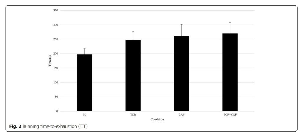
Theacrine also has an interesting role in cognition and inflammation. A paper using soccer players administered theacrine or theacrine plus caffeine to players and compared their performance in a simulated match. The study concluded that theacrine alone or theacrine plus caffeine improved performance, but theacrine plus caffeine was trending towards more improvement compared to theacrine alone. This study indicates that theacrine can enhance performance, but in combination with a low dose of caffeine has a superior impact on performance.

(Bello et al., 2019)

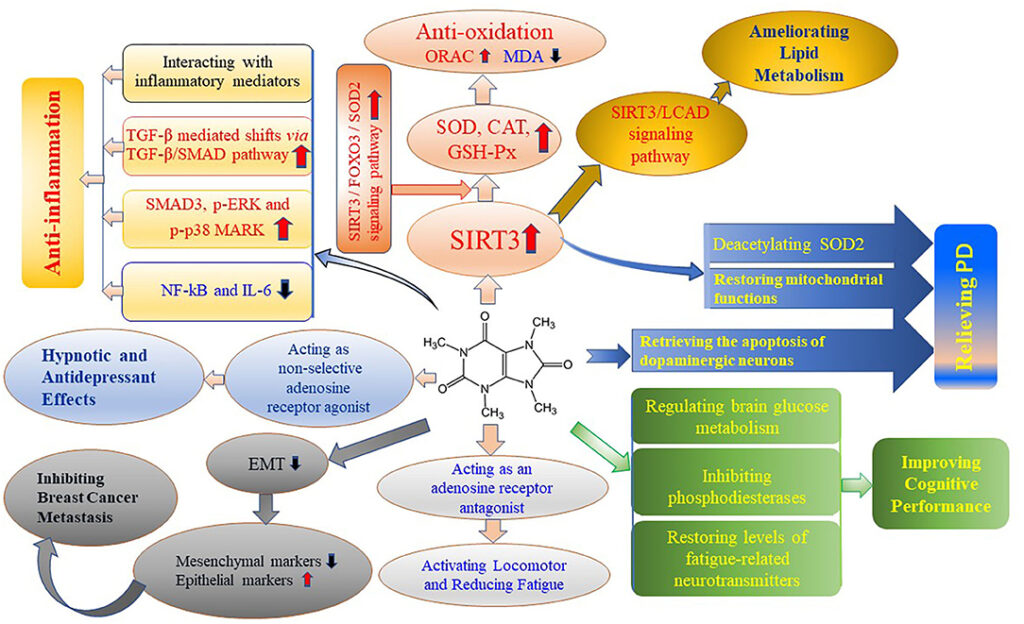
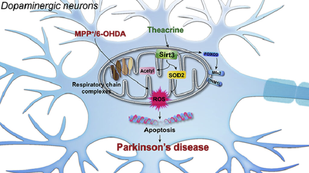
Theacrine’s impact on inflammation is a recent finding in the literature, with only a few studies to support its antioxidant capabilities. One of the first studies exploring theacrine’s antioxidant properties found that theacrine did not act directly to remove free radicals from the cell. Free radicals or reactive oxygen species can increase inflammation by causing damage to the cell, which activates a mechanism in the cell that causes apoptosis. There are several ways to increase these free radicals, such as chronic stress, trauma, or chemical exposure. Theacrine treatment of stressed mice, which induces a pro-inflammatory state, increased the expression of several genes involved in clearing reactive oxygen species and free radicals. The genes that theacrine upregulated were superoxide dismutase, glutathione peroxidase, and catalase, all of which act to decrease free radicals directly. A more recent study found that theacrine is able to activate sirtuin 3 (SIRT3), a member of the sirtuin family. SIRT3 is responsible for regulating apoptosis, or programmed cell death.

  • Apoptosis = Programmed cell death that results from cell damage or a signaling message. This cell death can cause inflammation due to the release of intracellular contents into the extracellular space.

SIRT3 regulates apoptosis through superoxide dismutase 2 (SOD2). SOD2 is a mitochondrial dismutase involved in clearing free radicals, thus promoting healthier mitochondrial health and preventing apoptosis. It was shown in the study that theacrine directly activates SIRT3, which protects against apoptotic processes. The big insights come from the fact that theacrine is able to cross the blood-brain barrier, which means the protective properties of theacrine can be facilitated in the brain. A study investigated Parkinson’s-induced mice, which notoriously have a loss of dopaminergic neurons and behavioral issues. The first conclusion the authors made was that theacrine treatment was able to improve behavior in Parkinsonian mice, suggesting the role of theacrine in the dopaminergic system. Next, the authors concluded that theacrine treatment alleviated dopaminergic neuron loss in zebrafish. The importance of this finding is that the death of a neuron is oftentimes a permanent loss due to the slow process of mitosis in neurons. In conclusion, theacrine offers protection from apoptosis through activating SIRT3, which downstream increases free radical scavengers and decreases pro-inflammatory processes.

Theacrine is a very useful supplement that has the capability to increase wakefulness, and productivity, act as an anti-inflammatory molecule, and enhance performance. Outside of its benefits, the single most important property of theacrine is that the body does not seem to build a tolerance, which suggests chronic use is safe and effective. This is important because, unlike caffeine, the dose of theacrine can stay the same over time, thus not inducing any toxicity-related side effects. Theacrine is effective at a wide range of doses; perhaps the most commonly used is 0.5–4 mg/kg. For a 170 lb (77 kg) male, an effective dose of theacrine is 100 mg.

Meet The Author

Hello everyone, 

My name is Joshua Giblin. I am a post-bachelor researcher/research technician at USC. My interests range from nutrition to nanomedicine and also practical science to improve everyday life. Through this blog, I aim to communicate practical scientific research and present it to curious individuals so that an educated decision can be made. Thank you for reading the blog and showing your support. 

Literature cited

  1. Bello, M. L., Walker, A. J., McFadden, B. A., Sanders, D. J., & Arent, S. M. (2019). The effects of TeaCrine® and caffeine on endurance and cognitive performance during a simulated match in high-level soccer players. Journal of the International Society of Sports Nutrition, 16(1), 20. https://doi.org/10.1186/s12970-019-0287-6
  2. Qiao, H., Ye, X., Bai, X., He, J., Li, T., Zhang, J., Zhang, W., & Xu, J. (2017). Theacrine: A purine alkaloid from Camellia assamica var. kucha with a hypnotic property via the adenosine system. Neuroscience Letters, 659, 48–53. https://doi.org/10.1016/j.neulet.2017.08.063
  3. Sheng, Y.-Y., Xiang, J., Wang, Z.-S., Jin, J., Wang, Y.-Q., Li, Q.-S., Li, D., Fang, Z.-T., Lu, J.-L., Ye, J.-H., Liang, Y.-R., & Zheng, X.-Q. (2020). Theacrine From Camellia kucha and Its Health Beneficial Effects. Frontiers in Nutrition, 7. https://www.frontiersin.org/articles/10.3389/fnut.2020.596823
  4. Taylor, L., Mumford, P., Roberts, M., Hayward, S., Mullins, J., Urbina, S., & Wilborn, C. (2016). Safety of TeaCrine®, a non-habituating, naturally-occurring purine alkaloid over eight weeks of continuous use. Journal of the International Society of Sports Nutrition, 13(1), 2. https://doi.org/10.1186/s12970-016-0113-3
  5. Wang, Y., Yang, X., Zheng, X., Li, J., Ye, C., & Song, X. (2010). Theacrine, a purine alkaloid with anti-inflammatory and analgesic activities. Fitoterapia, 81(6), 627–631. https://doi.org/10.1016/j.fitote.2010.03.008
  6. Ziegenfuss, T. N., Habowski, S. M., Sandrock, J. E., Kedia, A. W., Kerksick, C. M., & Lopez, H. L. (2017). A Two-Part Approach to Examine the Effects of Theacrine (TeaCrine®) Supplementation on Oxygen Consumption, Hemodynamic Responses, and Subjective Measures of Cognitive and Psychometric Parameters. Journal of Dietary Supplements, 14(1), 9–24. https://doi.org/10.1080/19390211.2016.1178678

Journal Review #9: The Positive Impacts of a High-Fat Diet on Synaptic Strength and Behavior

Background 
Depression and other mental illnesses are major problems in society. The cost of mental illnesses in the United States alone is closing in on $200 billion USD. Even with an enormous cost, many individuals are not receiving adequate assistance or reaching out to professionals, in part due to social norms and cost. Although there are many adults in the United States with depression, it may be relatively novel that these disorders stem from the adolescence period, and not necessarily the immediate time frame. The reason the adolescence periods are so crucial is that the brain and body are changing quite rapidly, and changes in hormones, growth factors, and nutrient intake all impact the brain. In turn, the brain can control the release of certain hormones as well. These feedback loops are crucial when examining the period of adolescence, which should be defined in this scenario as age 10 – 23 years old, partially due to the flexibility of the body at this age. 

A major environmental factor for proper development is nutrition. Evidence in the literature suggests that too steep of a calorie deficit can have negative impacts on long-term brain health. The literature also suggests that over-consumption of calories during adolescence is just as harmful, causing obesity, early onset diabetes, and inflammation. Until recently, the impact of a specific diet has not been known to impact brain structure. This alteration in brain structure can ultimately change behavior and increase the likelihood of depression and other mental illnesses.

Journal Review 
The aim of this article is to demonstrate the impact of a high-fat diet on brain structure and composition. Firstly, mice were taken at 2 months old and fed either a standard diet (SD) or a high-fat diet (HFD). After examining both diets’ compositions, it seems as if the HFD is calorically dense than the SD which may impact the results of this paper. This difference in calories had a significant impact on weight gain, with the HFD group gaining significantly more weight over the course of a month. With that being said, the difference in calories may or may not be significant, the researchers should have included this analysis in the research. Also, the conclusion from this paper should include the comparison between a high-calorie high-fat diet compared to a standard diet rather than a HFD versus a standard diet. 
It is also important to note that the brain is not composed of only neurons, there are many other cells that make up the brain. These cells have an important role in inflammation, protection, regulation, and metabolism. One of these cells is called astrocytes, a specialized glial cell involved in providing building blocks to neurons. These astrocytes are also involved in protecting neurons by providing glutathione precursors to neurons, with glutathione being the body’s main antioxidant. Although these astrocytes play a large role in many physiological processes, under specific environmental or genetic conditions, they can lead to pathophysiological disease states. This principle goes for many cells that have a role in regulating the immune system such as the different macrophage phenotypes, M1 and M2.

The importance of these astrocytes regarding dietary intake of fuel sources is regarding the mitochondria. The mitochondria house fuel from glycolysis, and use it to power the Krebs cycle, and electron transport chain, ultimately leading to the production of ATP. Since the cell can control the expression of genes through epigenetic modifications, it is not surprising that after time a change in diet changes the expression of metabolic genes. For example, a diet rich in carbohydrates may have higher expression of glycolysis genes, while a diet rich in fats may have higher expression of lipid utilization genes. In relevance to this journal article, the researchers found that the astrocytes in the hippocampus of the HFD mice had significantly higher amounts of lipids. The researchers also found higher amounts of c- and b-type cytochromes in the astrocytes of the HFD mice. However, when analyzing neurons, the HFD mice had no significant changes in lipid or cytochrome amount. From this data, the researchers concluded that a HFD induces metabolic changes in astrocytes not neurons in the hippocampus, which may affect the production of reactive oxygen species (ROS) present in the brain. 

Another aspect of astrocyte biology the researchers examined was morphology. To examine the size of astrocytes, hippocampal astrocytes were extracted and stained with dyes. After dying the components, a 3-D model can be constructed. After construction, the researchers concluded that astrocyte size in the hippocampus of HFD mice had increased branch length and domain area, which impacts the overall area that the astrocyte has action over. Next, the researchers used fluorescence to visualize the number of cells and the number of coupled cells in the hippocampus. The resulting images display that the HFD did not significantly affect the total number of cells nor the number of coupled cells in the hippocampus. 

Figure 2: 3-dimensional model illustrating astrocyte shape and size (A). Graph demonstrating number of intersections per astrocyte in the HFD or control model (B).
Figure 3: (D) Demonstration that the max number of intersections per astrocyte is significantly higher in astrocytes of the HFD group. (E) Graph illustrating mean branch length is significantly higher in HFD astrocytes relative to control astrocytes.

Figure 4: Astrocytes dyed with sulforhodamine 101 (SR101) in control and HFD groups. Imaged with fluorescent microscopy.

The researchers then focused on long-term potentiation (LTP), a process that strengthens the connection between synapses such that a long-term increase in neurotransmission occurs. It was found that long-term potentiation magnitude was significantly higher in HFD mice cells compared to control cells. The researchers then blocked the glutamate transporters using, TFB-TBOA, and repeated the previous experiment and found that field excitatory postsynaptic potentials (fEPSPs) magnitude, a measurement of LTP was no longer significantly different in HFD cells when compared to control cells. The result of this study is that the enhanced glutamate transport via HFD astrocytes may lead to enhanced LTP in the hippocampus. 

Figure 5: Graphs illustrating long-term potentiation via fEPSP.

To determine the impact of a HFD on behavior, the researchers performed an open-field test. The mice fed a HFD spent an equal amount of time moving, but a significantly higher amount of time in the center of the arena. Being in the center of the test indicates a lessening of anxiety. Therefore, the researchers concluded that a HFD led to an increase in synaptic potentiation in the hippocampus. This increase in hippocampal long-term potentiation led to a decrease in anxiety compared to mice fed a standard diet.

Figure 6: Graphs illustrating mice behavior.

Takeaways
There are several takeaways from this paper, including the possibility that a short-term increase in fat intake may have a beneficial impact on astrocyte physiology. These benefits may include decreasing anxiety through an increase in synaptic strengthening in the hippocampus. This study is a very fundamental paper and for the data to be reliably relayed to the public, the experiments need to be reproduced in a variety of environments.

Critiques
Keeping in mind that this study is a very basic paper, involving only a few data points in a specific brain region, the paper still has flaws that should be fixed in future studies. First, the caloric value of each diet should be properly gathered and used as a control, this way caloric variability does not influence the results. Secondly, more anxiety tests need to be run to make the conclusions the researchers make. The researchers use an open-field test to determine the impact of diet on anxiety, however, several tests should have been conducted to reproduce the results. 

Meet The Author

Hello everyone, 

My name is Joshua Giblin. I am a post-bachelor researcher/research technician at USC. My interests range from nutrition to nanomedicine and also practical science to improve everyday life. Through this blog, I aim to communicate practical scientific research and present it to curious individuals so that an educated decision can be made. Thank you for reading the blog and showing your support. 

Literature cited

  1. Firth, J., Gangwisch, J. E., Borsini, A., Wootton, R. E., & Mayer, E. A. (2020). Food and mood: How do diet and nutrition affect mental wellbeing? BMJ369, m2382. https://doi.org/10.1136/bmj.m2382
  2. Fu, W., & Jhamandas, J. H. (2020). Chapter 20—Amylin and amylin receptors in Alzheimer’s disease. In C. R. Martin & V. R. Preedy (Eds.), Genetics, Neurology, Behavior, and Diet in Dementia (pp. 309–324). Academic Press. https://doi.org/10.1016/B978-0-12-815868-5.00020-7
  3. Jha, M. K., Kim, J.-H., Song, G. J., Lee, W.-H., Lee, I.-K., Lee, H.-W., An, S. S. A., Kim, S., & Suk, K. (2018). Functional dissection of astrocyte-secreted proteins: Implications in brain health and diseases. Progress in Neurobiology162, 37–69. https://doi.org/10.1016/j.pneurobio.2017.12.003
  4. Popov, A., Brazhe, N., Fedotova, A., Tiaglik, A., Bychkov, M., Morozova, K., Brazhe, A., Aronov, D., Lyukmanova, E., Lazareva, N., Li, L., Ponimaskin, E., Verkhratsky, A., & Semyanov, A. (2022). A high-fat diet changes astrocytic metabolism to promote synaptic plasticity and behavior. Acta Physiologica236(1), e13847. https://doi.org/10.1111/apha.13847
  5. Sidoryk-Wegrzynowicz, M., Wegrzynowicz, M., Lee, E., Bowman, A. B., & Aschner, M. (2011). Role of Astrocytes in Brain Function and Disease. Toxicologic Pathology39(1), 115–123. https://doi.org/10.1177/0192623310385254

Supplements for Improving Neurite Growth and Membrane Integrity

Background
Maintaining neuronal membrane integrity is crucial for cognition. As humans age, mutations occur throughout our cells’ genetic material, resulting in a myriad of problems. One such problem is metabolism, in which transporters in our intestinal epithelial cells can no longer efficiently transport vital nutrients into the bloodstream. Another issue, independent of age, is dietary intake. An insufficient amount of a nutrient may negatively impact cognition due to the impact of the missing nutrient on cell integrity or neurotransmission. The last problem mentioned is the impact of chronically used pharmaceutical agents on cognition. A significant player in neuron death is excitotoxicity, which can result from too much excitatory signaling following the administration of a pharmaceutical agent. Pharmaceutical agents, especially ones where the dose is at the upper limit and usage is chronic, can alter gene expression in the neurons, resulting in tolerance, toxicity, and disruption of baseline levels of a gene even after use ceases. The problems previously mentioned call for a solution that aids in restoring neurotransmission and maintaining neuron integrity.  

Biology of Cell Membranes
There are several biology courses that focus on the fundamental components of the cell. Unfortunately, these lectures do a great disservice to demonstrating the importance of the cell membrane. As previously described, the cell membrane is involved in cell-to-cell recognition, cell signaling, enzymatic reactions, and separating the intracellular contents from the extracellular space. The cell membrane does not just desperate the spaces; it manipulates the environment and receives feedback from the molecules present. In relevance to brain health, the cell membrane of neurons is especially important because it is the hydrophobic nature of the membrane that allows neurotransmitters to be released into the synapse. These membranes are made of molecules called phosphatides, with the most abundant being phosphatidylcholine. 

(Structure of the Plasma Membrane (Khan Academy)

Biochemistry of Cell Membranes 
The formation of phosphatidylcholine is dependent on the reaction between choline and cytidine triphosphate to form cytidine diphosphocholine or CDP-choline. CDP-choline can then react with diacylglycerol, found in the cell membrane, to form phosphatidylcholine. Since this is a complex set of reactions, there must be enough precursor molecules, choline, cytidine triphosphate, and diacylglycerol, to adequately produce phosphatidylcholine. Since cytidine and uridine are both pyrimidines, an increase in circulating uridine will cause an increase in cytidine because they can be converted into one another rather easily. Since diacylglycerol is also a precursor to phosphatidylcholine, a polyunsaturated fatty acid such as docosahexaenoic acid (DHA) may be used to increase the levels of diacylglycerol when needed. 

(Cansev & Wurtman, 2007)

The Trifecta for Neuronal Membrane Integrity
Since the synthesis of phosphatidylcholine, a major player of the neuronal cell membrane requires choline, diacylglycerol, and cytidine triphosphate, supplements can be taken to indirectly increase phosphatidylcholine synthesis. First, a choline source is vital for phosphatidylcholine synthesis as well as acetylcholine synthesis. Secondly, uridine monophosphate (UMP) can be supplemented to indirectly increase the levels of cytidine triphosphate. Lastly, DHA, an omega-3 polyunsaturated fatty acid can be supplemented to increase levels of available diacylglycerol. Altogether, the three ingredients, choline, UMP, and DHA should be considered when maximizing neuronal membrane integrity. 

Choline
Choline is an essential nutrient with a recommended daily intake of 550 mg/day. More recent studies utilizing choline sources such as CDP-choline and alpha-GPC have used up to 1,200 mg/day with promising results and without any adverse effects. Alpha-GPC is reported to cross the blood-brain barrier more efficiently than other forms of choline and is able to raise acetylcholine levels in the hippocampus. A more recent 2022 paper found that alpha-GPC can enhance brain-derived neurotrophic factor (BDNF) and neuronal differentiation in the hippocampus. There are two papers in the last 5 years citing evidence that alpha-GPC may promote the formation of calcifications in the vasculature (atherosclerosis), so abuse of this supplement may not be warranted. The target dose for increasing acetylcholine via alpha-GPC is 300-600 mg/day. A later blog post will be analyzing the use of an alpha-GPC supplement as a cognitive enhancer. Although alpha-GPC is a valuable form of acetylcholine that promotes cognition-enhancing effects, CDP-choline is more valuable to this stack due to its immediate use in phosphatidylcholine synthesis. CDP-choline in this stack is useful at a dose of 100-600 mg/day with a majority of users finding that 200-400 mg/day works best. 

Effects of alpha-GPC supplementation on the novel object test after exposure to a non-stressor or stressor. (Jeong Yu et al., 2022).

Uridine Monophosphate
Uridine monophosphate acts as a reservoir for cytidine, such that uridine monophosphate can increase levels of cytidine triphosphate. This increase in cytidine triphosphate can lead to an increase in phosphatidylcholine synthesis. However, there are studies alluding to the fact that uridine monophosphate may also impact neurite formation through increased phosphatidylcholine. The first paper was conducted in vitro and demonstrated that uridine was able to increase the number of neurites, or neuron projections, compared to control cells. To determine the mechanism by which uridine monophosphate was increasing neurite abundance, cells were treated with apyrase. Apyrase is an enzyme that cleaves diphosphate and triphosphate nucleotide compounds, suggesting that UMP increases CTP levels, which can increase neuritogenesis. 

(A) In vitro assay demonstrated that incubating 50uM of UMP with a fixed number of neurons increased neurite outgrowth. (Pooler et al., 2005)
(B) 4 day incubation of UMP at 50uM was able to signficantly increase mean number of neurites per neuron.

Another paper looking at dopamine release found that uridine monophosphate administration increased striatal potassium-induced dopamine release. This paper also confirms that UMP treatment increases neurofilament-70 and neurofilament-M proteins, which are hallmark proteins involved in neuritogenesis. Although there are limited human studies, a 2011 trial found that UMP significantly decreased symptoms of depression. It should be mentioned that uridine monophosphate is not heavily researched, but the prevailing research seems to be extremely promising for future trials. Altogether, a dose of 100–500 mg/day is well tolerated in humans and demonstrates positive effects. There have been reports using up to 1,000 mg/day without any severe adverse effects. 

The effects of UMP supplementation on levels of neurofilament-70 (A) and neurofilament-M (B). (Wang et al., 2005)
UMP supplementation increases potassium-evoked dopamine release in the striatum. (Wang et al., 2005)

Docosahexaenoic Acid
Docosahexaenoic acid (DHA) is an omega-3 polyunsaturated fatty acid involved in a myriad of physiological processes. Although it is commonly found in fish oil, the recommended daily intake of 1,000 mg/day is rarely met in Americans. DHA has pronounced effects on inflammation and brain health. It is referred to as a pleiotropic molecule in the brain because it can assist structurally and impact neurotransmission. DHA is structurally important for phosphatidylcholine synthesis and cell membrane integrity. DHA supplementation when combined with uridine monophosphate administration was also found to have a positive effect on the restoration of dopaminergic neurons. A 2008 study investigated the effects of DHA and uridine supplementation on pre- and post-synaptic membranes, concluding that the combination was effective at increasing synaptic membrane number. A similar study using DHA and uridine found that synaptic membrane levels were significantly increased in the DHA + uridine group compared to control groups. Altogether, there are countless studies referencing the benefits of DHA on overall health, however, relevant to neuronal membrane integrity, DHA has a positive impact on synaptic membrane levels when taken with uridine. 

Impact of UMP plus DHA supplement on total phospholipids in both lesioned and intact striatum in a rodent model. (Cansev et al., 2008)

Impact on treatments on lesioned side of the striatum in a rodent model. The result indicated that UMP plus DHA supplementation significantly increased synapsin-1 levels, a protein involved in neuron formation. (Cansev et al., 2008)

Conclusions 
Maintaining neuronal membrane integrity is an ever-important function that requires a variety of precursors. Providing sources for the precursors in the forms of uridine monophosphate, choline, and Docosahexaenoic acid is useful for promoting membrane integrity. Along with the fundamental benefits that go along with membrane integrity, the combination of DHA and UMP increases the number of neurites or neuronal connections. Other reports have found that administration of UMP increases potassium-induced striatal dopamine release which can be useful when maximizing productivity. In conclusion, the combination of CDP-choline, UMP, and DHA provides an increase in phosphatidylcholine, the main component of the cell membrane which results in an increase in neurite projections and associated improvements in cognition. 


Meet the Author

Hello everyone, 

My name is Joshua Giblin. I am a post-bachelor researcher/research technician at USC. My interests range from nutrition to nanomedicine and also practical science to improve everyday life. Through this blog, I aim to communicate practical scientific research and present it to curious individuals so that an educated decision can be made. Thank you for reading the blog and showing your support. 

Literature cited

  1. Arenth, P. M., Russell, K. C., Ricker, J. H., & Zafonte, R. D. (2011). CDP-Choline as a Biological Supplement During Neurorecovery: A Focused Review. PM&R3(6, Supplement), S123–S131. https://doi.org/10.1016/j.pmrj.2011.03.012
  2. Cansev, M., Ulus, I. H., Wang, L., Maher, T. J., & Wurtman, R. J. (2008). Restorative effects of uridine plus docosahexaenoic acid in a rat model of Parkinson’s disease. Neuroscience Research62(3), 206–209. https://doi.org/10.1016/j.neures.2008.07.005
  3. Cansev, M., & Wurtman, R. J. (2007). Chronic administration of docosahexaenoic acid or eicosapentaenoic acid, but not arachidonic acid, alone or in combination with uridine, increases brain phosphatide and synaptic protein levels in gerbils. Neuroscience148(2), 421–431. https://doi.org/10.1016/j.neuroscience.2007.06.016
  4. Jeengar, M. K., Thummuri, D., Magnusson, M., Naidu, V. G. M., & Uppugunduri, S. (2017). Uridine Ameliorates Dextran Sulfate Sodium (DSS)-Induced Colitis in Mice. Scientific Reports7(1), 3924. https://doi.org/10.1038/s41598-017-04041-9
  5. Jeong Yu, H., Lin Kim, Y., Jung Kim, M., Mee Park, J., Young Park, S., Nae Park, S., & Won Yang, D. (2022). The effect of choline alphoscerate on non spatial memory and neuronal differentiation in a rat model of dual stress. Brain Research1786, 147900. https://doi.org/10.1016/j.brainres.2022.147900
  6. Kondo, D. G., Sung, Y.-H., Hellem, T. L., Delmastro, K. K., Jeong, E.-K., Kim, N., Shi, X., & Renshaw, P. F. (2011). Open-Label Uridine for Treatment of Depressed Adolescents with Bipolar Disorder. Journal of Child and Adolescent Psychopharmacology21(2), 171–175. https://doi.org/10.1089/cap.2010.0054
  7. Li, B., Zhou, H., Wu, X., Chen, Z., Yao, J., & Yin, Y. (2016). Effects of dietary supplementation with uridine monophosphate on performance and intestinal morphology of weanling piglets1. Journal of Animal Science94(suppl_3), 82–86. https://doi.org/10.2527/jas.2015-9440
  8. Lopez, C. M., Govoni, S., Battaini, F., Bergamaschi, S., Longoni, A., Giaroni, C., & Trabucchi, M. (1991). Effect of a new cognition enhancer, alpha-glycerylphosphorylcholine, on scopolamine-induced amnesia and brain acetylcholine. Pharmacology Biochemistry and Behavior39(4), 835–840. https://doi.org/10.1016/0091-3057(91)90040-9
  9. Nutrients | Free Full-Text | DHA Effects in Brain Development and Function. (n.d.). Retrieved September 13, 2022, from https://www.mdpi.com/2072-6643/8/1/6
  10. Ocalan, B., Cakir, A., Koc, C., Suyen, G. G., & Kahveci, N. (2019). Uridine treatment prevents REM sleep deprivation-induced learning and memory impairment. Neuroscience Research148, 42–48. https://doi.org/10.1016/j.neures.2019.01.003
  11. Pooler, A. M., Guez, D. H., Benedictus, R., & Wurtman, R. J. (2005). Uridine enhances neurite outgrowth in nerve growth factor-differentiated pheochromocytoma cells. Neuroscience134(1), 207–214. https://doi.org/10.1016/j.neuroscience.2005.03.050
  12. Structure of the plasma membrane (article). (n.d.). Khan Academy. Retrieved September 15, 2022, from https://www.khanacademy.org/science/ap-biology/cell-structure-and-function/plasma-membranes/a/structure-of-the-plasma-membrane
  13. Tuboly, E., Gáspár, R., Ibor, M. O., Gömöri, K., Kiss, B., Strifler, G., Hartmann, P., Ferdinandy, P., Bartekova, M., Boros, M., & Görbe, A. (2019). l-Alpha-glycerylphosphorylcholine can be cytoprotective or cytotoxic in neonatal rat cardiac myocytes: A double-edged sword phenomenon. Molecular and Cellular Biochemistry460(1), 195–203. https://doi.org/10.1007/s11010-019-03580-1
  14. Uridine Ameliorates Dextran Sulfate Sodium (DSS)-Induced Colitis in Mice | Scientific Reports. (n.d.). Retrieved September 13, 2022, from https://www.nature.com/articles/s41598-017-04041-9
  15. Wang, L., Pooler, A. M., Albrecht, M. A., & Wurtman, R. J. (2005). Dietary Uridine-5’-Monophosphate Supplementation Increases Potassium-Evoked Dopamine Release and Promotes Neurite Outgrowth in Aged Rats. Journal of Molecular Neuroscience27(1), 137–146. https://doi.org/10.1385/JMN:27:1:137
  16. Wang, Z., Hazen, J., Jia, X., Org, E., Zhao, Y., Osborn, L. J., Nimer, N., Buffa, J., Culley, M. K., Krajcik, D., van den Born, B.-J. H., Zwinderman, K., Levison, B. S., Nieuwdorp, M., Lusis, A. J., DiDonato, J. A., & Hazen, S. L. (2021). The Nutritional Supplement L-Alpha Glycerylphosphorylcholine Promotes Atherosclerosis. International Journal of Molecular Sciences22(24), 13477. https://doi.org/10.3390/ijms222413477