Uridine monophosphate (UMP) is a nucleotide, which is a building block of RNA (ribonucleic acid). UMP is synthesized in the liver and is also found in dietary sources such as mushrooms, broccoli, and organ meats. UMP has been found to play a role in a variety of biological processes, including the biosynthesis of RNA and DNA, as well as in the regulation of various neurotransmitters in the brain. UMP supplementation has been studied for its potential cognitive enhancing effects and is commonly used as a supplement to improve memory and focus.
In addition, uridine is a precursor to several important brain chemicals, including phosphatidylcholine, which is a major component of cell membranes and is involved in cell signaling. Phosphatidylcholine is also a precursor to acetylcholine, a neurotransmitter involved in learning and memory. By supplementing with uridine monophosphate we can improve several aspects with positively impact our productivity, including neurogenesis (growth of neurons) and acetylcholine signaling.
Mechanism of Action
Uridine monophosphate (UMP) is bioavailable and can be taken orally as a supplement. When ingested, it can be converted to uridine and incorporated into the body’s total nucleotide pool, which can support various cellular processes. UMP can also cross the blood-brain barrier, allowing it to affect brain function directly.
Since RNA codes for proteins, by increasing the amount of nucleotides for RNA synthesis, we can upregulate several processes in the cell. This upregulation can lead to increased dopamine signaling and cell growth.
UMP also acts as a precursor to phosphatidylcholine (PC), the predominant lipid constituting our cell membranes. Since neurogenesis, the growth of neurons, depends on the cell going through mitosis, the cell needs to increase the amount of available PC, so it can successfully split into two distinct neurons.
Figure 1. Mitosis.
Evidence from the Literature
There is a lack of evidence using UMP supplementation to improve aspects of memory or mood in humans. However, this should not steer us away from investigating its use as an aid in our daily life.
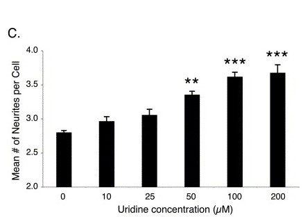
One paper investigated the effects of uridine monophosphate (UMP) on neuronal outgrowth in primary cultured neurons. The results showed that UMP significantly increased neurite length, the number of neurites, and the number of branch points in neurons. UMP treatment also increased the expression of genes related to neurite growth, including BDNF, NGF, and GAP-43. The authors concluded UMP has a positive effect on neuronal outgrowth.
Another study investigated the effects of uridine supplementation on learning and memory in rats subjected to hypobaric hypoxia, which can cause neuronal death and impair cognitive function. The researchers found that uridine supplementation improved learning and memory in the rats, and protected against hypoxia-induced neuronal death in the hippocampus. The study also suggested that uridine may enhance mitochondrial function in the hippocampus, which could contribute to its neuroprotective effects.
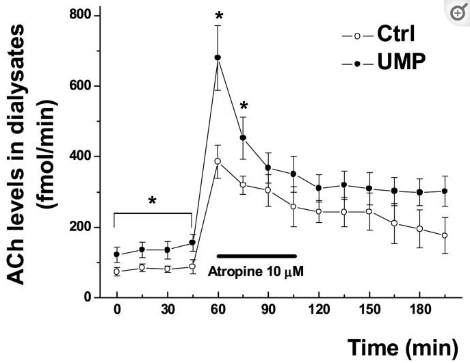
The last study I will mention aimed to investigate the effect of dietary supplementation with uridine-5′-monophosphate (UMP) on the acetylcholine level and release in the striatum of aged rats. The researchers found that the dietary supplementation of UMP significantly increased the levels of acetylcholine in the striatum of aged rats. This increase was also associated with enhanced release of acetylcholine. The results suggest that UMP supplementation can improve acetylcholine-related cognitive functions in aged rats.
Uridine has been shown to improve many cellular functions attributing to enhanced productivity. Anecdotal evidence suggests supplementation with uridine monophosphate at 100-400 milligrams can feel stimulating, similar to caffeine. The effects of UMP supplementation take place around 15 minutes post-ingestion and can last around 2-4 hours depending on previous nootropic usage, sensitivity, and experience.
As always, initial dosing protocols should be at the lower end of the recommended range. As for UMP, this should be at 50-100 milligrams. Subsequent doses can be tapered until a dose of 300-400 milligrams is reached, where the maximum effects can be felt.
There is limited research on the long-term safety of UMP supplementation. However, short-term studies suggest UMP is safe and well-tolerated, with no serious adverse effects reported. It is always recommended to consult with a healthcare provider before starting any new supplement, especially for long-term use. It is important to follow recommended dosages and not exceed them.
Concluding Remarks
The evidence suggests that uridine monophosphate (UMP) supplementation can enhance productivity through multiple mechanisms, such as improving cognitive function, increasing acetylcholine levels, promoting neuronal outgrowth, and enhancing mitochondrial function. While the evidence is still limited, the results from animal and human studies are promising and warrant further investigation. UMP supplementation appears to be safe and well-tolerated, making it a potential option for those looking to improve their productivity.
PFAs stand for Per- and Polyfluoroalkyl Substances, which are a group of man-made chemicals that have been used in a wide range of industrial and consumer applications for several decades. They were first introduced in the 1940s and have been used in a wide range of industrial and consumer applications since then. Initially developed for products such as non-stick cookware, waterproof clothing, and stain-resistant fabrics because of their unique properties of being resistant to heat, water, and oil. Since then, PFAs have been incorporated into thousands of products, including food packaging and electronic devices, leading to daily exposure, which may have devastating consequences.
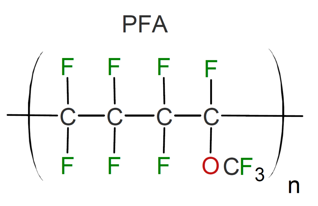
Figure 1. Image of Perfluoroalkoxy alkane.
PFAs are typically referred to as a double-edged sword. On one side, they are excellent materials for cookware, safety, and lab use because of their persistent properties. On the other side, their persistent means accumulation in the body which can have detrimental effects on our health. Some examples are that PFA is linked with decreased immune function, liver toxicity, and can increase the risk of cancer. These concerns warrant the increasing awareness of PFA’s danger to the environment and government interventions for limiting exposure.
PFAs and Health
The “forever chemicals” as you can guess, are chemicals that are very resistant to natural decomposition. PFAs are resistant to decomposition because of their unique chemical properties. They are composed of long chains of carbon and fluorine atoms, which make them very stable and resistant to breaking down in the environment. The carbon-fluorine bonds in PFAs are among the strongest chemical bonds found in nature.
The resilient nature of these chemicals makes it hard for our bodies to remove them, thus they accumulate in our organs and cause damage.
PFAs are linked to a range of health problems. The scientific community has been studying the health effects of PFAs for several years, and evidence suggests that exposure to these chemicals can have harmful effects on human health.
Some studies have linked PFAs to an increased risk of certain types of cancer, including kidney and testicular cancer. Exposure to PFAs has been linked to effects on the immune system, including decreased antibody response to vaccines, and developmental effects on fetuses and infants, including low birth weight and delayed development. PFAs may also interfere with hormones, which can have effects on reproductive and metabolic health.
Several studies have linked increased PFA exposure to liver damage, including a study published in the journal Environmental Health Perspectives in 2017 found that exposure to PFAs was associated with liver injury in a human population. The study found that individuals with higher levels of PFAs in their blood had increased levels of liver enzymes, which can show liver injury. Other studies have showed that increased exposure to PFAs leads to liver damage in rodent/zebrafish models.
How can you Limit Exposure to PFAs?
There are several ways to limit your exposure to PFAs. This list is not exhaustive nor erases your exposure to the forever chemicals, and perhaps the best way to limit PFAs exposure is to push for policy reform in your region. Currently, the United States Environmental Protection Agency (EPA) has established a health advisory level of 70 parts per trillion (ppt) for PFOA and PFOS (two types of PFAs) in drinking water. Several states have established their own regulations, including California and New Jersey, which have set maximum contaminant levels for PFAs in drinking water. These increasingly stringent regulations help protect citizens from the devastating effects of PFAs, but there is more work to be done. While you are pushing for policy reform, here are some tips for reducing your exposure to PFAs.
While it may seem obvious, steering clear from items that contain PFAs is the best way to start. Some of these items include non-stick cookware, waterproof clothing, stain-resistant fabrics, and food packaging.
Stainless steel or cast iron cookware can be used instead of non-stick cookware
Another tip is to avoid drinking water contaminated with PFAs. While government regulations have become increasingly more stringent, it is beneficial to get a reverse osmosis water system.
Reverse osmosis is an effective method for removing many contaminants, including PFAs, from drinking water. Reverse osmosis works by forcing water through a semipermeable membrane, which has a pore size of around 0.0001 microns, that filters out impurities, including PFAs, as the water passes through.
However, it is important to note that not all reverse osmosis systems are created equal, and some may be more effective than others at removing PFAs. Some types of PFAs may be more difficult to remove than others, and the effectiveness of reverse osmosis may depend on the specific type of PFAs present in the water.
The last tip for avoiding excess exposure to PFAs is to avoid certain seafoods.
According to the U.S. Food and Drug Administration (FDA), large predatory fish such as shark, swordfish, and tuna can accumulate higher levels of PFAs in their tissues. This is because they eat smaller fish that have also accumulated PFAs. Also farmed fish like salmon and trout, may be fed with fishmeal or fish oil that has been contaminated with PFAs. As a result, they may contain higher levels of PFAs than wild-caught fish.
Looking Forward
At a brief glance, it may be overwhelming to consider all the detrimental effects of PFAs on human health. Compounding PFAs impact together with the increasing abundance of microplastics and no one would blame you for becoming pessimistic about the future. Before you are totally convinced that your children and grandchildren will be permanently altered by the choices made by the previous generations, you have a choice in limiting your personal exposure to PFAs and having a positive impact on communities that do not receive the same attention. Help yourself and other by helping to reform policy around microplastics and PFAs in the environment.
Meet the Author
Hello everyone,
My name is Joshua Giblin. I am a post-bachelor researcher/research technician at USC. My interests range from nutrition to nanomedicine and also practical science to improve everyday life. Through this blog, I aim to communicate practical scientific research and present it to curious individuals so that an educated decision can be made. Thank you for reading the blog and showing your support.
Weight loss is fundamentally dependent on two variables. Calories being consumed and calories being expended. To lose weight, the number of calories being consumed must be less than the number of calories being expended. This is what we call being in a calorie deficit. Because the equation relies on two variables, we can manipulate each of them separately or together to create the calorie deficit. Namely, we can decrease calorie consumption by decreasing food intake, increase calorie expenditure by exercising, or decrease food intake and perform regular exercise.
Recent studies have concluded that combining a solid cardiovascular and resistance training program with a healthy diet improves metabolic parameters, longevity, and led to successful weight loss. For clients with limited to no experience dieting or losing weight, I recommend they spend 1-6 months optimizing their diet before starting a regular exercise program. This is because it can be tricky to change only your diet, so it can be extremely overwhelming to change both diet and add a new habit into your life. The 1-6 month window gives the client time to make dieting a habit, thus when they are ready to go to the gym, they are less stressed and able to make more out of the experience.
In order to compound diet and training atop each other, you must build a strong foundation in either. It is like building a house on sand. You may be able to live in it for a few months, but upon arrival of the first storm, your house will be swept away. Building a strong foundation when dieting is dependent on sustainability, nutrient intake, and tracking your progress.
Sustainability
As discussed in The Weight Loss Journey, we know that steady weight loss decreases the likelihood of binging episodes. The sustainability aspect of losing weight allows for prolonged weight loss, better maintenance after the weight loss phase, and improved skills in building better habits. However, this is easier said than done. Building a diet that contains important macro/micronutrients goes against the typical western diet,
often rich in saturated fats, processed foods, and simple sugars. Not only is the switch in diet socially difficult, it is also psychologically difficult. Going against the mainstream media and having to isolate your environment can be detrimental to your psychology, which can lead to a buildup of emotions and eventually a binging episode. Going with this, modern western culture is flooded with fast food on every corner and processed calorie-dense foods all over the grocery store. Fortunately, there are practical changes that can help make your diet more sustainable.
The first approach is to maximize food volume. A common mistake is confusing food volume with food calories. For example, eating one pint of ice cream can be over 1000 calories, whereas the same volume of a vegetable may be a maximum of 200 calories. In this scenario, you are physically eating the same food volume (mass/amount) but by eating the vegetable; you are drastically reducing your caloric intake.
Now that you know food volume does not equate to caloric value, you can structure your diet to include at least one high-volume food item per meal. For example, if you aim to eat 350 calories for meal one, it may look something like this:
Food Item
Calories
Volume Rating
3 Large Eggs
210
Low
200g Egg Whites
104
Very High
4oz Spinach
30
Very High
In this example, there are 2 high volume foods, namely egg whites and spinach. This meal also has a good distribution of fats (eggs), protein (egg whites), and vitamin K (spinach). For those who enjoy eating carbohydrates in the morning, an example may look like this:
Food Item
Calories
Volume Rating
2 Large Eggs
140
Low
200g Egg Whites
104
High
4oz Spinach
30
Very High
1/2 Cup Oatmeal
150
Medium
Including the oatmeal adds valuable carbohydrates and other nutrients while still being a low calorie meal by excluding 1 egg. Both meals are great options, however, mixing and matching food preferences are a necessity for sustainability. If you don’t enjoy eating eggs, find a food similar in terms of nutrients or calories that you enjoy eating.
Building a sustainable diet for weight loss can be tricky, and it can often take months to find a subset of healthy foods you enjoy. After learning about how to manipulate food volume, the next step is tracking your daily food intake so we can clearly see patterns.
Tracking Your Food
Tracking your food intake is important for several reasons. First, it acts as an accountability agent, making it more likely for you to adhere to your diet. Second, it allows for long-term pattern analysis of your diet. You can see how your diet has developed. Lastly, it can provide valuable insight into micro/macronutrient intake, both of which are important for health. For example, if you are eating lots of dairy products one week and are having gastrointestinal issues, then you can clearly correlate the problem. If you were not tracking your food, it can be easily forgotten hence leading to a longer time dealing with the issue, resulting in mental stress and increasing your likelihood of failure.
Perhaps the easiest method for tracking your food intake is by utilizing an app like cronometer. Cronometer is a free app that tracks your food and analyzes your nutrient intake.
Nutrient Intake
The last step in building a sturdy foundation upon which you can start building a better diet is in regards to nutrient intake. A nutrient is a substance that is needed to promote growth and maintenance. Simply put, you need nutrients to live. However, just receiving the bare minimum is not optimal for health. A lack of nutrients can lead to several different diseases but more commonly slight deficiencies may promote unhealthy mechanisms in the body. For example, a lack of vitamin K can promote an osteopathic state. It is crucial to receive nutrients at proper doses, especially while dieting since our bodies will be running on fewer calories. Therefore, those calories must be nutrient-dense to promote fat loss and health.
There are several classes of nutrients. Perhaps the more popular are macronutrients. These are simply nutrients needed in abundance (grams). The most commonly known macronutrients are protein, carbohydrates, fats, and fiber.
Protein
Protein intake should be around 1.6-1.8g/kg body weight, for example, a 165lb male (74.8kg) should be eating 120-135 grams of protein per day. Protein is important for the maintenance of skeletal muscle, cell integrity, growth, and repair. Good sources of protein are lean cuts of animal meat, namely, chicken breast, ground beef, and steak. Other sources include peas, egg whites, and milk.
Fats
Fats are crucial for many physiological and anatomical reasons. Some of the most important molecules in our bodies, hormones, are made from cholesterol. Without cholesterol, testosterone and estradiol levels would be crushed. Fats are also important for energy storage and insulation. A great place to start is 35% of your total calories coming from fats. Great sources of fat include eggs, sunflower seeds, animal meats, and extra virgin olive oil.
Carbohydrates and Fiber
Carbohydrates are extremely important for the body. They act primarily as fuel sources. Complex carbohydrates can be broken down by digestive enzymes into glucose, a direct fuel source. Carbohydrates are also used in cell signaling and recognition. Good carbohydrate sources include oatmeal, sweet potatoes, fruits, and beans. It may be important to eat carbs in a timely manner, shuttling carbohydrates closer to the evening. The purpose of doing this is based solely on cognitive performance. Meals higher in carbs may negatively affect productivity.
Fiber is an important micronutrient. It provides fuel for your macrobiota and promotes gut health. Good sources of fiber include oats, sweet potato, beans, and some fruit. One of the most beneficial supplements, psyllium husk, is a perfect source of fiber.
Micronutrients
As opposed to macronutrients, micronutrients are substances needed in small amounts, think milligrams, not grams. Some popular examples are magnesium, B vitamins, vitamin D, potassium, and sodium. Maintaining micronutrient intake is especially important during weight loss because food intake is low. This decrease in food intake leads to fewer micronutrients being consumed. This is why choosing the right foods is crucial.
Micronutrient
Food Source
Amount from Food
Recommended Daily Intake (RDI)
Vitamin K
4oz Spinach
547 ug
120 ug
Magnesium
30g Chia Seeds
117 mg
400 mg
Potassium
100g banana
358 mg
3400 mg
Selenium
15g Brazil Nuts
479 ug
55 ug
Zinc
4oz Ground Beef
5.6 mg
11 mg
Vitamins B3 & B6
4oz Chicken Breast
14.1 mg B3 0.7 mg B6
16.0 mg B3 1.3 mg B6
Key Takeaways
When building your diet for weight loss, it is important to account for several factors. Some of these factors include sustainability and adequate nutrient intake. Sustainability in your diet is important. Being able to lose weight over time is crucial for your short- and long-term health. Your diet should also be rich in micronutrients like zinc, iron, and the b vitamins to maintain optimal metabolism and performance. Along with eating micronutrient rich foods, properly balancing macronutrients like protein, carbohydrates, and fats is equally important. Eating enough protein allows for muscle maintenance, carbohydrates for fuel, and fats for proper hormone production. Of course, different ratios exist between these macronutrients and finding which works the best for you will take time.
For personalized nutrition or training programs, feel free to email me at [email protected].
Meet the Author
Hello everyone,
My name is Joshua Giblin. I am a post-bachelor researcher/research technician at USC. My interests range from nutrition to nanomedicine and also practical science to improve everyday life. Through this blog, I aim to communicate practical scientific research and present it to curious individuals so that an educated decision can be made. Thank you for reading the blog and showing your support.
Losing weight at its fundamental core revolves around thermodynamics. Namely, that energy cannot be created or destroyed, rather, it can be transferred from one form to another. A plant receiving lots of energy from sunlight can transfer that energy into storage substances. If the plant is releasing half of that energy in order to maintain structure, it is storing the other half. Over time, this leads to a surplus of stored energy. This same fundamental principle applies to humans. If more energy from food (calories) is being consumed than is being released, an excess of storage will occur. The primary storage for humans being glycogen and triacylglycerol (TAG), where glycogen is found in skeletal muscle and liver, and TAG is found in adipose (fat) tissue. Glycogen storage is quickly maxed out if excess calories are being consumed, leaving the rest of the energy to be stored in fat tissue.
If you aim to lose weight, you must consume fewer calories than you expend. This is commonly termed as being in a calorie deficit. Another easy example is found in finances. If you are making $10.00 /day but you are spending $12.00/day, you would lose $2.00/day. To compensate, you tap into your savings account, thus depleting your savings. The opposite would occur if you made $10.00/day but spent only $8.00/day. This would net you $2.00/day in profits that need to be stored in the bank. In this example, the dollar amounts represent calories, while the bank represents fat storage. In short, to lose weight, you must consume fewer calories than you expend, leaving your body with no choice but to tap into its storage.
Know Your Measurements
In order to be in this caloric deficit, you need to know how much food you can eat. If you are not eating enough, your metabolism will crash, micronutrients will become scarce, and you will feel like crap. You will eventually hit a massive plateau in your weight loss journey. There are several methods to optimize weight loss. The most efficient is using a Calorie Counter. This method accounts for gender, activity, height, and weight to give you an estimated number of calories your body needs to maintain its size. For example, a 22-year-old male, standing 5 feet 6 inches, exercising 5 times per week, needs 2500 calories to maintain weight. To lose weight, we can subtract from this maintenance number, thus putting us in a caloric deficit. While this part of the journey seems trivial, it provides a target, that being an achievable number, which is much more sustainable than winging it.
The next step is choosing how many calories to subtract from our maintenance. You may see people subtracting 1,000 calories from their baseline. Subsequently, you will witness these same people binging during the weekend, hitting plateaus, and being miserable. A sensible approach would be to subtract 350 calories from maintenance. Under perfect conditions, not accounting for water weight, this would result in weight loss at a rate of 1 lbs per 10 days. Not only is taking away 350 calories going to grant you substantial weight loss, it will also be sustainable. The process also reserves muscle and will be much more enjoyable than crash dieting.
Now that we know that weight loss occurs when our body is expending more than it is consuming, we have subtracted 350 calories from our maintenance. By doing so, we can enjoy steady weight loss while limiting episodes of binge eating and misery.
Track Your Progress
Congratulations, we are losing weight at a sustainable pace, now we need progress markers to properly map out the journey. Think of tracking your progress as a way of checking the fuel in your car. Starting with a full tank, you can drive away. However, if you don’t check the fuel gauge, before you know it, you’re empty. Stuck on the side of the highway, waiting for aid. We can prevent this by checking the fuel gauge. In the same sense, we can check our weight loss gauge. Unlike a car, whose miles per gallon is relatively the same whether the fuel tank is full or low, the body adapts. As you decrease calories, metabolism slows down, and over time, what was your losing weight calorie number is now your maintenance calories number. Now, weight loss is halted, but if you didn’t track your weight, you would be stuck. Luckily, you tracked your weight using a scale, and you can see that after X weeks of eating 350 fewer calories than your maintenance, weight loss has slowed or halted.
Tracking your weight is relatively easy. Not only does it take only 20 seconds, but it is extremely cheap and valuable. Tracking your weight daily is preferred. It can be at anytime; however, keeping food/water intake consistent before weighing is important. If one day you drank 50oz of water before stepping on the scale, you would weight 1.4kg more because of the water. This wouldn’t be an issue, except people rarely drink water at the same time every day, besides upon waking. Therefore, taking your weight upon waking is the most efficient. It not only gives you the lowest weight of the day, but the most consistent measurement.
Fortunately, you have been tracking your progress and have lost significant weight. This is amazing, but now for the past 3 weeks, you have seen no drop in weight, and are left wondering where to go from here.
Hitting A Plateau
When battling with weight loss, it can be demoralizing to hit a plateau. Then again, now that you understand what it means, we can start losing weight in no time. A plateau is when weight loss slows to almost none, and it happens because the body adapts to the amount of calories you have been feeding it. It does this by primarily decreasing heat output. Now that we know our body’s new maintenance calorie is lower than previously discussed, we have three main options. One, we can decrease caloric intake by another 350 calories, therefore reinstating a caloric deficit and burning fat. Two, we can increase energy output by exercising. I plan to cover exercise in an upcoming article. However, performing resistance training 3-6 times per week, coupled with 120 minutes of cardiovascular exercise, will increase your energy expenditure by a substantial amount. This increase will tip the scales, leaving you in a caloric deficit and back to losing weight. The last option includes a blend of one and two. A slight calorie reduction, possibly 100-300, and introducing exercise in conjunction. The scientific literature heavily supports the notion that exercise increases health and lifespan, making it a vital addition to your life. I am adding exercise to the option list when the first plateau is reached because eating healthy is a large enough task to begin with. It may overwhelm someone trying to learn both exercise and diet, and that feeling of stress increases the likelihood of failing. At this point in your journey, you may know the foods you enjoy. You may have built this diet into a habit, so now is a perfect opportunity to add exercise.
After learning what a weight loss plateau means for your journey, you have a few choices to make. Decrease current calories by another 350, add exercise, or the most efficient choice, slightly decreasing your calories by 100-300 and adding exercise to your routine.
A Major Blockade
You may have hit a few plateaus since we last spoke, as expected. Great, you applied the options given to you in the previous section and got back to losing weight. Now you find yourself, perhaps several months/a year deep in the diet, and you feel as if your weight loss has stagnated. Before despair settles, this is very predictable. As time passed, you kept decreasing your caloric intake and increasing exercise with every plateau. At some point, a blockade hit. This is the point where you can’t lower your calories without feeling terrible or being nutrient deficient, and you can’t do more exercise. This blockade, if dieting is done properly, should not be encountered for 6 months to 2 years, depending on your level at the start of this journey.
The Restoration
While there are several options, crash dieting or lowering your calories beyond your tolerability are not some of them. Crash dieting will exponentially increase the likelihood of a binge episode and weight regain. It is crucial at this stage to implement a metabolism restoration period. This period relies on slowly ramping up your metabolism by increasing caloric intake in small increments. This will cause weight gain that is controlled, desired, and beneficial to your long-term health as opposed to crash dieting.
To start metabolic restoration, start by bumping up your caloric intake by 200-400. This will ensure that your metabolism will ramp up, while still not allowing your body to store too many calories as fat. With the previous condition, these calories should be made of protein (80%), which acts to reduce the bacteria species responsible for lipid absorption, thus limiting fat accumulation at this stage.
The restoration period can be thought of as changing the oil in your car. As you drive, oil is used up, eventually leading to low oil levels. These low oil levels can negatively affect your car’s lifespan and performance, therefore, you fill the oil and clear up the filters. Similarly, oil can represent food. As you have dieted, food intake has steadily decreased, eventually reaching a blockade, where food intake is low enough to affect performance. To combat this, bump up the food in a controlled manner and get those energy levels back up.
A New Beginning
After you have raised your calories by 200-400, your weight in the next few weeks will reach its new maintenance weight. Depending on your weight loss goals, this may be your ideal body fat level, whereas others may have their eyes set on losing a little more fat. For those who have reached their new ideal body fat, congratulations are in order. While the maintenance phase is still difficult and requires mindful eating, by now, habits like eating healthy foods, performing regular exercise, and appreciating your body dominate the mind.
For those looking to lose a bit more body fat, this restoration period serves as the ultimate test. When a crash dieter has no experience with maintaining a level of body fat, they lose control, binge, and regain lots of fat. However, you know better. Repeatedly ramping up your calories slowly until a new maintenance weight is reached allows for complete control of weight gain. This increase in calories, specifically protein, will grant increased muscle accrual, thus further improving your metabolic health.
After several rounds of restoration, largely based on several factors, including performance and happiness, the weight loss journey can resume.
The Final Stretch
After months or years of improving your mental and physical health, you have made it to the final stretch. As you now know, in order to lose weight, you must be in a caloric deficit, which can be achieved by increasing exercise or decreasing caloric (food) intake. With the restoration period resulting in a new maintenance calorie amount, which should have been properly tracked, start by subtracting 100-300 calories. As you can see, the last stretch does not differ from what you have already mastered before. The same rules apply, eat slightly less, perform exercise, and reap the rewards.
Hitting the Mark
The process of switching between restoration and caloric deficit until you have reached your ideal body fat is quite subjective. Often, what someone wants is not ideal for their body and will ultimately end in disappointment and episodes of binge eating. The standard seen on tv is fake. A facade made of makeup, CGI, angles, extreme crash dieting, and often the result of performance-enhancing drug use (steroids). At this point in your journey, the ideal body fat for you lies between psychological satisfaction and sustainability. If you repeatedly lose fat and then uncontrollably gain it back, this is a sign that your psychological satisfaction is not matched with your sustainable body fat. This will be different for everyone, and is often a learned process that may take years to find out, since it may be psychologically rooted.
Hitting the mark means setting a realistic body weight, being both satisfactory and sustainable. Of course, this does not mean your journey is over. Over the course of the past year, training hard in the gym has become a habit, allowing you to gain more muscle, lose fat if warranted, and feel at your best, and this does not change now that you have reached your ideal body weight.
Continuous Progress
Congratulations, you have arrived at what you deem is the closest point in time to your goal body. This body, merely a vehicle for your now improved lifespan, has helped craft habits like eating healthy and going to the gym. Yet your body does not define who you are. You are more than the person who lost weight. You are an entrepreneur, a parent, a sibling, a hard worker, a fit person, and the list goes on. Do not let the completion of a goal like weight loss stop you from crafting better habits, whether they are big or small.
You have amassed skills like training in the gym and eating healthy to overcome your biological precedent and improve your life.
For many readers, you may only be a few weeks into your lifestyle change, making it difficult to imagine a time where you can continuously overcome problems. Let this serve as a message that momentum in the positive direction and in all aspects of life acts as a bullet-proof vest. Your future self will face the same problems you do today. They just have a thicker vest on through years of successful habit building.
Concluding Remarks
Losing weight is a laborious task. It involves going against your biological processes and reversing decades of weight gain. The reason no timeline is mentioned is simply that people proceed at different speeds. Maybe subtracting 100 calories from your diet was difficult, resulting in an increase of time spent dieting. As intended, this article is not a one-size fits all, but a general template to follow and it lays out the future obstacles with the answers. It is like receiving a pre-filled out study guide for an exam. After reading this, when you inevitably hit a plateau, you will know what to do.
Other articles, with a similar style, tackling different obstacles in the weight loss journey will be posted. Some of these posts focus on…
Nutritious foods to eat while dieting
A guide to creating a sustainable diet
Resistance training and why it is important
With all of this being said, thank you for reading. I am optimistic and believe that you can accomplish this goal. For coaching, email me at [email protected]
Meet the Author
Hello everyone,
My name is Joshua Giblin. I am a post-bachelor researcher/research technician at USC. My interests range from nutrition to nanomedicine and also practical science to improve everyday life. Through this blog, I aim to communicate practical scientific research and present it to curious individuals so that an educated decision can be made. Thank you for reading the blog and showing your support.
Part 1: Prevent Excess Fat Accumulation after Dieting
New Year New You
As the 2022 holiday season concludes, a long list of goals for 2023 start to gain traction. Among them, losing weight is both popular yet extremely difficult. It takes months to adjust your appetite and pick up valuable training information, and it takes even longer for the results to appear. With all the hardships that come with losing weight, it still remains the best way to increase your lifespan and quality of life.
Perhaps the most frustrating aspect of the weight loss journey is the post-diet weight gain. While dieting, we restrict calories below that of caloric expenditure, resulting in a caloric deficit. Forcing your body to burn fat cells for fuel. To the average person, this is weight loss on the surface. However, after several months of dieting, calories must be continuously decreased to account for metabolic homeostatic processes, thus leading to a plateau in the weight loss journey. While at the plateau stage, it is oftentimes recommended to increase calories slightly, therefore, allowing the body to increase metabolism and caloric expenditure. During this post-diet phase, weight gain can be extremely discouraging to anyone and often results in uncontrollable eating and weight gain, thus reverting to progress back to none.
This “Yo-Yo” dieting is seen as a lack of discipline or motivation, thus casting yet another shadow of hate onto an individual who’s in a biological war. In fact, there are several hormonal factors at play which impacts people differently. Therefore, a healthy body weight is the body weight most sustainable while still not increasing risk for disease. For example, person A has great health, found by medical examinations, but has a BMI of 25. According to classic BMI, it deemed this person overweight, yet when person A tries to lose fat, they end up starving, not enjoying life, and eventually binge eating. In this scenario, person A while dieting is miserable, less social, and creates a health catastrophe in binge eating. At this point, person A is healthy at a BMI slightly above the universal standard.
This example is relatable for many people. However, getting blood work and medical examinations to test your health is required. If you need to lose weight because of your health decline, dieting comes in all shapes and sizes. Be sure to research the best ways to limit hunger or find a professional to optimize weight loss. Also, being under the guidance of an accountability coach can help make the journey more efficient. Learn more about coaching here.
Keep the Weight Off
To prevent sudden weight gain during a plateau period, a diet high in protein is great practice. Not only will protein consumption significantly improve muscle maintenance during the dieting phase, but prevents excess fat accumulation during the plateau/re-feeding stage. A recent article published in naturemetabolism highlights the importance of a high protein diet and preventing fat accumulation after a period of caloric restriction.
To briefly highlight the mechanism revealed in the keystone article, they fed mice a high protein diet after a period of caloric restriction. Resulting in significantly less fat when calories were increased to back to baseline compared to mice fed a variety of diets. The authors showed that the high protein diet prevents fat accumulation via inhibiting/preventing Lactobacillus growth, a species of bacteria responsible for intestinal lipid absorption. By preventing lipid absorption, fewer calories are absorbed into the bloodstream, preventing excess fat accumulation.
This study contains practical information, such that a high protein diet is beneficial when trying to maintain weight loss resulting from a short period of caloric restriction.
Proper protein consumption can be very tricky. First, tracking nutrition is vital to monitoring your health. Apps like cronometer make this first step extremely simple. By putting in all of your foods, you can see which vitamins, minerals, and macronutrients you are missing. This is vital for tracking protein consumption because too much protein may negatively impact longevity while too little protein will cause muscle loss and other problems. 1.6 grams of protein per kilogram of body weight seems to be an appropriate amount of protein that does not sacrifice longevity nor muscle mass. A 200 pound (90.7 kg) male would need to take in 144 grams of protein to meet this recommendation.
Meet the Author
Hello everyone,
My name is Joshua Giblin. I am a post-bachelor researcher/research technician at USC. My interests range from nutrition to nanomedicine and also practical science to improve everyday life. Through this blog, I aim to communicate practical scientific research and present it to curious individuals so that an educated decision can be made. Thank you for reading the blog and showing your support.
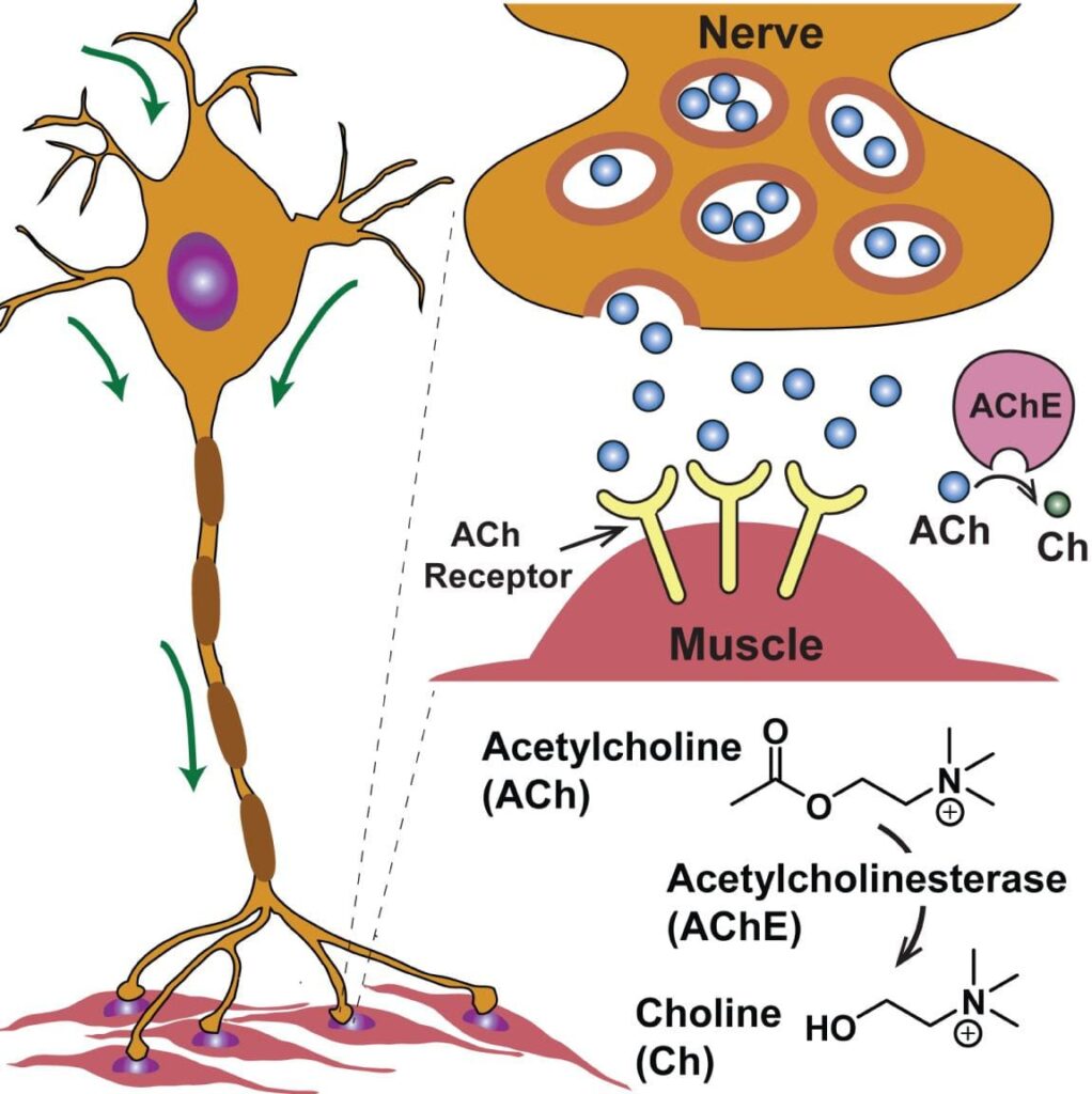
Neurotransmission and regulation of neurotransmitter release are key components in regulating behavior. Of the neurotransmitters, acetylcholine, also a neuromodulator, plays a crucial role in attention, behavioral control, and muscle contraction. Chemically, acetylcholine requires choline and acetyl-CoA for synthesis, and is released by cholinergic neurons throughout the brain and at neuromuscular junctions. Acetylcholine binds to the muscarinic or nicotinic cholinergic receptor, each receptor family containing multiple subtypes, all with varying functionality. Adequate acetylcholine regulation is critical for attention, as shown in rats that displayed significant impairments in attention when treated with a nicotinic acetylcholine receptor (nAChR) antagonist. Beyond attention, acetylcholine is critical for proper inhibitory control, an executive function that allows for inhibition of habitual behaviors. However, the data pooled from rodent and human studies regarding acetylcholine and inhibitory control have been quite contradicting. This current review from a paper published in Scientific reports on December 3rd, 2022, titled Acetylcholine deficit causes dysfunctional inhibitory control in an aging-dependent manner, offers a model for analyzing acetylcholine’s action on inhibitory control in drosophila.
Takeaways: Acetylcholine, a neuromodulator, is assembled from choline and acetyl-CoA. Acetylcholine is released by cholinergic neurons, found throughout the brain, and plays a vital role in inhibitory control, attention, and muscular contraction. Due to contradicting results when comparing rodent and human models of acetylcholine’s impact on inhibitory control, this journal review explores a fly model.
Results
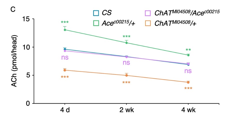
Inhibitory control is essential for executing goal-directed behavior. Improper regulation of inhibitory control leads to impulsive actions and thoughts, thus deterring the subject from goals that are not yet habitual. To assess the change in inhibitory control over-time, the researchers used a go/no-go test (GNG). The GNG test measures constraint against a no-go signal. Under no wind conditions, flies are free to move about the cage. However, introduction of a strong wind results in a decrease in movement, most likely to increase survival percentage during a strong wind event. To test whether age increased the number of inhibition events, the authors counted how many times the fly disobeyed this inhibitory behavior, thus notating this action “loss of inhibition events” (LIE) at different time points. To implicate the impact of acetylcholine, the researchers compared wild-type (CS) to Ace-c00215 flies. The Ace-c00215 allele is a null variant encoding acetylcholinesterase, therefore there is significantly less acetylcholinesterase in these flies compared to wild-type flies. The less acetylcholinesterase is present, the more acetylcholine is active in the synaptic cleft.
The first figure [1B] relays information about the total number of LIE in 4d, 2wk, and 4wk old flies that have a wild-type or the Ace/+ genotype. Two primary results can be concluded from this figure. First, regardless of acetylcholinesterase level, the number of LIE increases in an age-dependent fashion. Second, the increase of acetylcholinesterase (Ace) significantly reduces the number of LIE compared to wild-type (CS) flies. This result suggests a large role for acetylcholine in inhibitory control.
Figure 1B
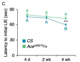
The second figure [1C] compares the latency to the initial LIE in wild-type (CS) and Ace flies over-time. An increase in latency to the initial LIE shows positive inhibitory control. The wild-type flies show a significant decrease in latency time to initial LIE when compared to Ace flies, further implementing acetylcholine’s role in inhibitory control. The decrease in acetylcholinesterase prevents the decrease in latency time to initial LIE in flies up to 4 weeks.
Figure 1C
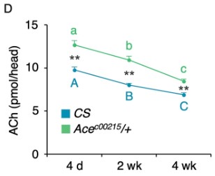
The authors measured acetylcholine (ACh) levels as seen in figure [1D]. In both wild-type (CS) and Ace flies, acetylcholine levels decline over-time, suggesting an age-dependent decrease in acetylcholine. Also, the Ace flies show significantly more acetylcholine at each time point compared to wild-type mice. Together, these results show acetylcholine declines in an age-dependent manner, as well as implements acetylcholine as a key player in inhibitory control.
Figure 1D
Takeways: The loss of inhibition events (LIE) refers to an event in which a fly moves during an inappropriate time. The total number of loss of inhibition events increases in an age-dependent manner and is significantly reduced in flies with increased acetylcholine. The results implement acetylcholine as a regulator of inhibitory control, and acetylcholine decreases over-time, indicating an increase in loss inhibition events as flies age.
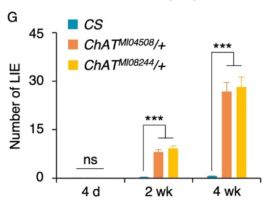
To further support the notion that acetylcholine is vital for inhibitory control, the researchers genetically removed the choline acetyltransferase (ChAT) enzyme. ChAT is the enzyme responsible for the biosynthesis of acetylcholine from choline. The ChAT enzyme is encoded by one gene in both humans and drosophila, making this a practical model. Using an immunoreactivity test, the researchers verified the ChAT M104508 and ChAT M108244 heterozygous variants had significantly less acetylcholine compared to wild-type flies (CS) [Figure 2B, C].
After verifying successful ChAT suppression, the researchers used the same experiment as previously described to determine the number of LIE. Briefly, flies were exposed to strong wind conditions, and movement was deemed a LIE. The results [Figure 2G] compared the total number of LIE over-time in 1) ChAT M104508 2) ChAT M108244 or 3) wild-type (3) flies. At 4 days, there are no significant differences between treatment groups. However, in an age-dependent manner, the total number of LIE increases in either of the ChAT groups. The results from this experiment suggest a crucial role for acetylcholine in inhibitory control, such that acetylcholine-deficit impairs inhibitory control.
Figure 2G
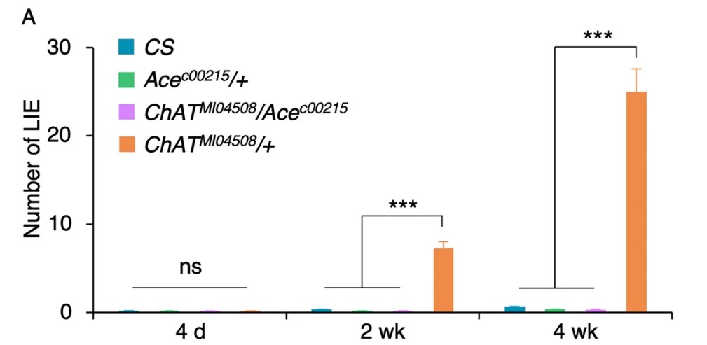
To verify the role of acetylcholine in inhibitory control, the researchers compared a variety of genetically differing flies. The genetic make-up of the groups are: 1) Ace-c00215/+ 2)ChAT-MI04508/+ 3) ChAT-MI4508/+ / Ace-c00215 and 4) wild-type (CS). The Ace groups contain a mutation that blunts acetylcholinesterase, thus increasing acetylcholine. Whereas, the ChAT groups harbor a mutation resulting in a decrease in choline acetyltransferase, thus decreasing acetylcholine. As seen in figure [3A], the total number of LIE significantly increases over-time in all groups, this verifying an age-dependant function in inhibitory control. Also, the addition of the Ace mutation alleviates the impact of the ChAT mutation on the number of LIE.
Figure 3A
The researcher quantified the amount of acetylcholine in each treatment group. As expected, the ChAT mutation resulted in a significant decline in acetylcholine levels compared to all other groups. Also, the ChAT/Ace group showed wild-type levels of acetylcholine, thus supporting the conclusion from [3A]. Of note, the group harboring the Ace mutation had significantly increased acetylcholine level at any age point.
Figure 3C
Discussion
Acetylcholine dysregulation has been connected with several diseases, including Alzheimer’s disease. It modulates a large population of neurons and has critical roles in attention. Currently, its role regarding inhibitory control, an executive function allowing for inhibition of a habitual action, has been under-fire due to contradicting results in human and rodent models. The aim of this paper was to identify the role of acetylcholine in inhibitory control using genetically modified drosophila or fruit flies. Flies harboring a mutation resulting in decreasing acetylcholinesterase showed significantly better inhibitory control compared to wile-type flies. To further support the role of acetylcholine in inhibitory control, flies containing a null choline acetyltransferase allele showed significantly worse inhibitory control compared to wild-type flies. To verify the findings, the authors showed that flies harboring both mutations (increased acetylcholinesterase/ decreased choline acetyltransferase) had restored inhibitory control and increased acetylcholine levels compared with choline acetyltransferase mutated flies. The researchers also identified that this inhibitory control decreases with age along with levels of acetylcholine.
Acetylcholine deficit results in significantly impaired inhibitory control
Practical Takeaways
Inhibitory control refers to the ability to not perform a behavior that is deemed natural or habitual to complete a goal. This is also referred to as impulse control. Key ideas around success stem from delaying instant gratification. For example, hunger on a diet is near impossible to curve without abusing stimulants or pharmaceuticals. The ability to sustain a caloric-deficit and overcome the hunger is a positive action for the long-term goal of losing weight but goes against fundamental survival behaviors. Other examples, like watching television or over-consuming social media when other goals, whether financial, physical, or mental, are present, represent a lack of impulse control. The flooding of dopamine from social media is rewarding, while studying may instantly feel rewarding (without bio hacking). This paper alludes to the fundamental role of acetylcholine in inhibitory control, suggesting that increasing acetylcholine may lessen the number of impulsive actions. There also seems to be an age-dependent decrease in acetylcholine, hence, a decrease in the ability to control impulses. Possible remedies to control impulses may revolve around administration of alpha-GPC, which have showed to increase acetylcholine contents in the brain. These results suggest that dose-adjustments are needed in an age-dependent manner, with older individuals needing a higher dose.
Meet the Author
Hello everyone,
My name is Joshua Giblin. I am a post-bachelor researcher/research technician at USC. My interests range from nutrition to nanomedicine and also practical science to improve everyday life. Through this blog, I aim to communicate practical scientific research and present it to curious individuals so that an educated decision can be made. Thank you for reading the blog and showing your support.
Literature cited
Sabandal, P. R., Saldes, E. B., & Han, K.-A. (2022). Acetylcholine deficit causes dysfunctional inhibitory control in an aging-dependent manner. Scientific Reports, 12(1), Article 1. https://doi.org/10.1038/s41598-022-25402-z
Careful monitoring and adjustment of nutrient intake is among one of the most effective treatments for a variety of metabolic disorders, including obesity. Unfortunately, a majority of individuals enter a state of self-destruction by creating a non-sustainable and nutrient-deficient plan. These destructive actions are then further encouraged by many online influencers, promoting drastic non-scientific methods. These self-destructive patterns and increased food availability have ultimately led to a rise in obesity prevalence throughout most of the developed world, which has resulted in the obesity being the most prominent chronic disease. Besides lifestyle changes, bariatric surgery, the most preferred for patients, has been drastically improved over the years, yet still carries significant risks that can be mitigated through other measures. These measures include personalized nutrition and the use of pharmaceutical aids, with GLP-1 agonists and dual GLP-1/GIP agonists having tremendous therapeutic potential.
This journal review aims at presenting a study that deploys tirzepatide, a dual agonist of the glucagon-like peptide 1 (GLP-1) and glucose-dependent insulinotropic polypeptide (GIP) receptors. Before we discuss the therapeutic efficacy, a brief review of the GLP-1, GIP, and glucose regulation is warranted.
Glucose & Insulin
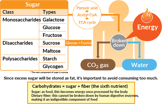
Glucose is the body’s primary fuel source, acting as a necessary molecule for glycolysis which fuels the Kreb’s cycle, and downstream cellular respiration. The intake of simple sugars can be quickly absorbed into the bloodstream, however, complex carbohydrates such as starch, which can be composed of multiple glucose molecules bound together. These complex carbohydrates must be broken down by amylases, released by the pancreas into the small intestine. Once the complex carbohydrates are broken down into the base molecule, glucose, it can be taken into the bloodstream and used by cells for energy. There are many complex systems involved in regulating blood glucose levels, including the release and mechanism of insulin.
As glucose molecules circulate, they enter through a glucose transporter on pancreatic beta cells, which results in the release of insulin. The role of insulin is to bind to the insulin receptor on target cells, which activates a plethora of signaling cascades, one of which is to insert glucose transporters into the cell membrane (GLUT). The GLUT allows the glucose from the bloodstream to enter cells and be used for energy, and without the GLUT protein, glucose cannot be used for fuel. Therefore, insulin is crucial for glucose regulation and cellular energy metabolism, thus its dysregulation is apparent in diseases like diabetes.
Takeaways: Carbohydrates from foods are broken down into their primary components, glucose. Glucose is a major source of fuel for the production of energy, and the consumption of glucose stimulates the release of insulin. Insulin’s role in the body is to decrease blood glucose by allowing cellular uptake of glucose.
When glucose is abundant, insulin stimulates cells to store glucose as glycogen in the liver and skeletal muscle. This stored glucose is incredibly useful for quick energy when glucose consumption is not abundant, such as periods of caloric-deprivation or exercise. During these periods, insulin release is inhibited by the actions of glucagon, released from pancreatic alpha cells. Glucagon’s main role as a hormone is to increase blood glucose when levels are too low, such as during fasting or exercise. To achieve glucose homeostasis, glucagon stimulates the liver to breakdown glycogen, thus releasing glucose into the bloodstream.
Takeaway: When blood glucose levels are low, glucagon is released which acts to inhibit insulin release and increase the breakdown of liver glycogen. The primary role of glucagon is to increase blood glucose levels.
Hormones & Digestion
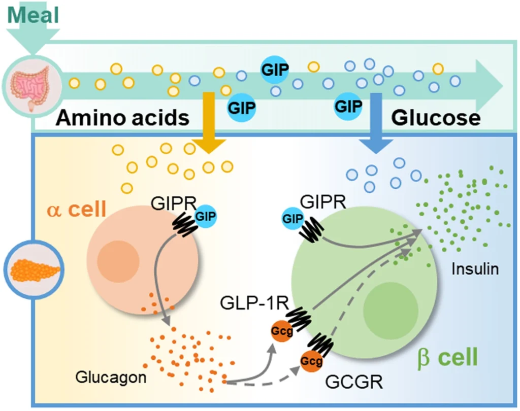
The digestive system is extremely complex, involving a variety of signals originating from the nervous and endocrine systems. Several hormones play a crucial role in regulating glucose, including, as previously mentioned, insulin and glucagon. However, other hormones offer a unique set of characteristics regarding glucose homeostasis, including glucagon-like peptide 1 (GLP-1) and glucose-dependant insulinotropic polypeptide (GIP).
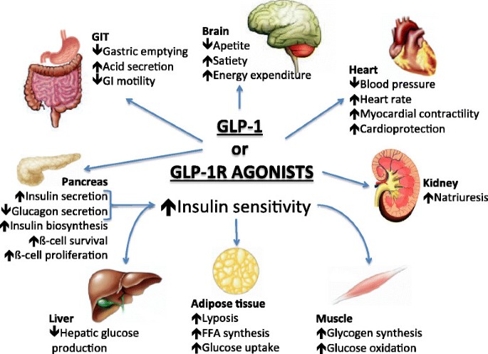
Endogenous GLP-1 is released from cells of the intestine when glucose is sensed and acts primarily to increase insulin secretion, increase insulin sensitivity, and delay gastric emptying. Exogenous GLP-1 receptor agonists exhibit the same effects as endogenous GLP-1, however, modifications can be made to increase time-of-action, thus lowering safety concerns around administration.
Endogenous GIP results in similar physiological changes compared to GLP-1. This includes an increase in insulin secretion and an increase in glucose usage. This hormone also acts to lower blood glucose by stimulating insulin release. GIP also plays a role in altering energy metabolism in the brain, thus making the combination of GLP-1 and GIP favorable for weight loss and treatment of type II diabetes. Exogenous GIP exhibits similar effects as endogenous GIP.
Takeaways: The digestive system is a complex system with a plethora of hormones that regulate satiety, glucose, and gastric motility. Among them, GLP-1 and GIP are important for satiety and glucose homeostasis through insulin release, satiety, and altering cell metabolism.
Obesity
As the prevalence of obesity continues to rise, the number of realized factors involved with the onset also increases. Obesity is characterized by having a body-mass index of above 30 mg/km, or a significantly high percentage of body fat accumulation. At a fundamental level, this is caused by an energy imbalance in which energy intake is greater than energy expenditure. As research continues to improve, genetics, psychology, environment, and hormone imbalances seem to play a large role in the development of obesity. This disease results in dysregulated eating patterns, increased adipose (fat) tissue, increased blood glucose, and increased insulin resistance. Each of these problems has a subset of issues, including an increase in pro-inflammatory fat-secreted hormones and thickened blood from increased glucose levels. To date, the treatment of obesity has been through behavioral clinics and pharmaceutical stimulants, which cause side effects that limit sustained use. Surgical intervention has also been used, with many patients reporting undesired side effects along with a significant price tag. Therefore, a pharmaceutical aide aimed at regulating several pathways, including behavioral and glucose homeostasis, is a favorable therapeutic option in treating obesity.
Takeaways: Obesity is a complex disease involving multi-faceted dysregulation. Hallmark features of obesity include a significant accumulation of adipose tissue along with increase blood glucose levels due to increased insulin resistance. Treatments centered on altering behavior and glucose levels are a promising therapeutic agent.
Tirzepatide Phase 3 Results
Methods
This clinical trial assessed the impact of tirzepatide, a dual GLP-1 and GIP receptor agonist, in obese patients with no history of diabetes. All participants had a body-mass index >30, no history of diabetes, no surgical intervention, and must have reported a previous unsuccessful dietary intervention prior to recruitment. These strict criteria allow the authors to present valid data, since diabetes can strongly influence the action of this drug, and surgical intervention may cause exaggerated side effects.
This phase 3 double-blind, randomized, controlled trial consisted of 2539 participants receiving once-weekly, subcutaneous tirzepatide at 5 mg, 10 mg, or 15 mg or placebo for 72 weeks. To assess the change in various markers, baseline measurements were taken at week 0 and compared to measurements taken at week 72.
Results
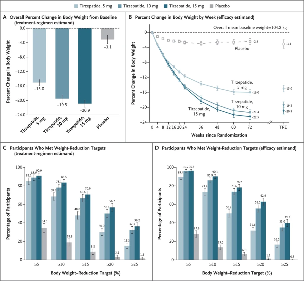
The results from this trial are promising for the future of treating obesity. First, all dosage groups receiving tirzepatide experienced a reduction of body-weight with the 15 mg group having the most substantial change in being -20.9%. The 5 mg group displayed -15.0 and the 10 mg group a -19.5 percent change in body weight. There seems to be no significant difference between the 15 mg and 10 mg groups, signifying a plateau in dose-response at around 10 mg.
Along with a significant change in percent body weight from baseline, the following graphs give insight into future dosing regimens. In figure B, the percent change in body weight by week contains a significant drop in weight by all tirzepatide groups from weeks 0 to 12. However, the 10 mg and 15 mg tirzepatide groups displayed a longer period of substantial weight loss, being from weeks 0 to 48 and afterwards plateauing.
Critical Analysis: A potential use for this therapeutic may deploy 10-15 mg for 48-60 weeks, followed by a rest period of 12-24 weeks for resensitization.
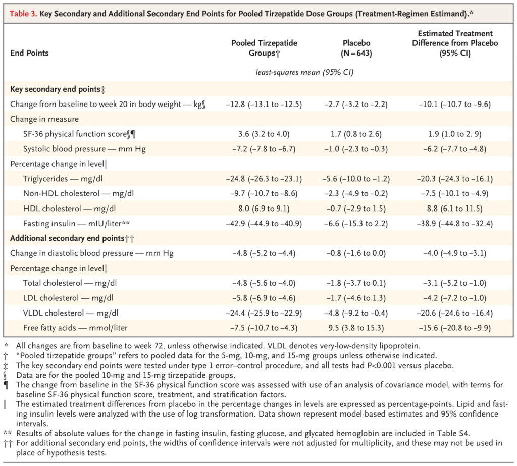
Other data points such as waist circumference, blood pressure, and fasting insulin were favorable after treatment with tirzepatide. Regarding diabetes treatment potential, the tirzepatide group exhibited significantly lower fasting insulin levels, dropping nearly 43% from baseline, with the placebo group only exhibiting a 6.6% decrease from baseline. Other data points that should have been considered for glucose regulation are fasting glucose levels and HbA1c at week 72.
Positive changes in low-density lipoprotein (LDL), high-density lipoprotein (HDL), free fatty acids, and systolic blood pressure were observed in the tirzepatide groups. These measurements, when pooled with the significant decrease in percent change in body weight, indicate that tirzepatide at 10-15 mg is an effective treatment for obesity, although a large population study must be performed to confirm its safety.
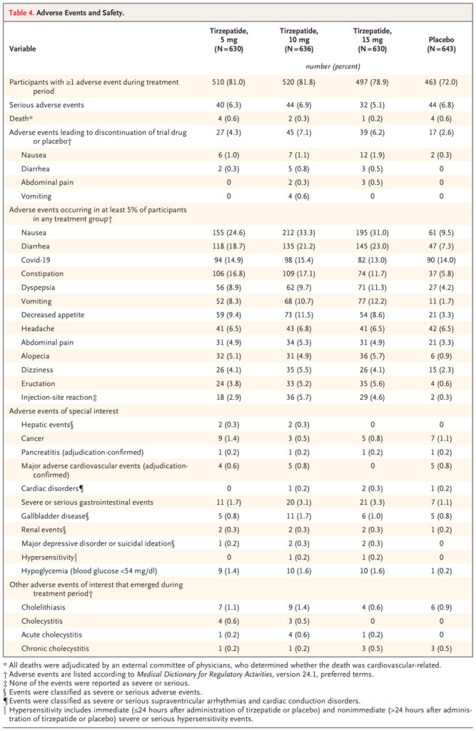
Takeaways:Tirzepatide at 10 or 15 mg once-weekly for 72 weeks resulted in a -19.5 or -20.9 percent change in body weight. The usage of tirzepatide also resulted in positive changes regarding LDL, HDL, and systolic blood pressure. The tirzepatide-groups had significantly more reported transient-to-mild adverse effects signifying the dosing protocol had an adequate safety profile.
Discussion
Obesity is the most prominent chronic disease in the world, resulting in an increased risk of cardiovascular disease and diabetes. Unfortunately, treatments have been centered on changing lifestyle habits, which is not a sustainable or realistic ideology for treating a multi-faceted disease involving many feedback loops. Tirzepatide, a peptide with dual agonist activity at the GLP-1 and GIP receptors, exhibits tremendous therapeutic potential. Over the course of 72 weeks, patients receiving a once-weekly subcutaneous injection of tirzepatide at 15 mg exhibited a 20.9 percent decrease in body weight change and positive changes in fasting insulin and lipid profile. The reported adverse effects were primarily transient-to-mild, thus supporting its use as a therapeutic peptide for the treatment of obesity.
Meet the Author
Hello everyone,
My name is Joshua Giblin. I am a post-bachelor researcher/research technician at USC. My interests range from nutrition to nanomedicine and also practical science to improve everyday life. Through this blog, I aim to communicate practical scientific research and present it to curious individuals so that an educated decision can be made. Thank you for reading the blog and showing your support.
Literature cited
Jastreboff, A. M., Aronne, L. J., Ahmad, N. N., Wharton, S., Connery, L., Alves, B., Kiyosue, A., Zhang, S., Liu, B., Bunck, M. C., & Stefanski, A. (2022). Tirzepatide Once Weekly for the Treatment of Obesity. New England Journal of Medicine, 387(3), 205–216. https://doi.org/10.1056/NEJMoa2206038
Plastics are durable, waterproof, and extremely cheap, making the exponential rise in use and production inevitable. With great demand in fields such as medicine, packaging, and electronics, the amount of plastic waste accumulating in the United States alone exceeds 40 million metric tons. Even with astounding advances in recycling engineering, the plastic recovery rate is less than 5%, meaning that almost all plastic waste is piled in the ocean and landfills.
As rain, wind, and mechanical erosion by dirt and other substances weather the plastics, it begins to slowly degrade. This process releases microplastics into the dirt and water, thus allowing humans to be exposed to these substances at abnormally high concentrations. The term microplastics refers to any plastic debris with a size of less than 5mm and can be further broken down into primary and secondary microplastic subcategories. Primary microplastics are purposefully designed for medicine and care products, while secondary microplastics are those small plastic particle debris resulting from decomposing plastic.
These microplastics cannot be filtered or removed from the environment by commercially available protocols and therefore have been accumulating uncontrollably in soil, water, and food sources for decades. Earlier studies have conducted the impact of micro- and nano-plastics in rodent models and human cells in vitro. In this brief article, the impact of microplastics on health, sources of microplastics, and a future guide regarding limiting microplastic exposure will be discussed.
Impact of Microplastics on Living Organisms
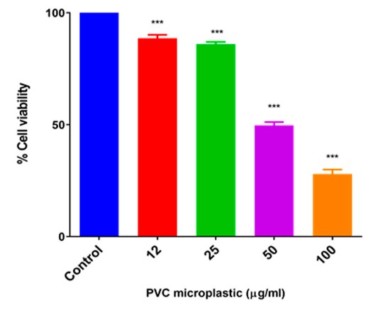
The accumulation of microplastics results in an increase in microplastic exposure, thus resulting in a higher microplastic burden on the body. As microplastics accumulate in the body, typical excretion methods become overburdened and immune cells may become activated. These immune cells can help eliminate the microplastics but inflammation can arise as a result. It is also seen throughout the literature that microplastics can enter the cell and result in decreased cell viability via DNA damage when the concentration of microplastics reaches as little as 12 ug/ml.
As seen in this figure, as the concentration of microplastics increases, the cell viability of human lymphocytes decreases. Cell viability refers to survival, and PVC is polyvinyl chloride, a type of microplastic. The significance of this work resides in the impact of microplastics on lymphocytes, which are immune cells.
Since an increase in microplastic concentration kills more lymphocytes, the resulting exposure of lymphocytes to microplastics creates a more susceptible immune system. More specifically, lymphocytes are a class of immune cells that houses B cells, T cells, and natural killer cells. These immune cells are crucial in protecting the body from bacteria, cancer, and viral infections.
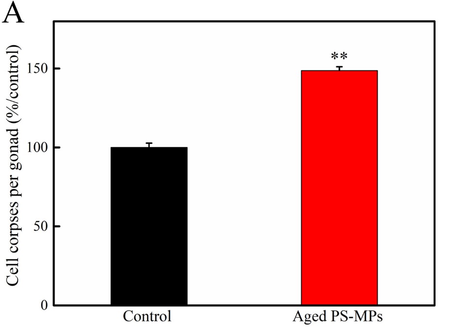
Certain microplastics are directly harmful to specific regions in the body such as reproductive organs. A 2022 study found that polystyrene microplastics (PS-MPs) exposed to UV light resulted in reproductive toxicity in C. elegans.
The first experiment examined the number of cell corpses, or dead cells, per gonad after exposure to UV-exposed PS-MPs. The results indicate that UV-exposed PS-MPs induced cellular apoptosis at a higher rate than gonadal cells not exposed to PS-MPs, suggesting that these microplastics are indeed toxic to reproductive organs.
The results from this paper confirm that PS-MPs exposed to UV light result in DNA damage which leads to cell death in reproductive organs.
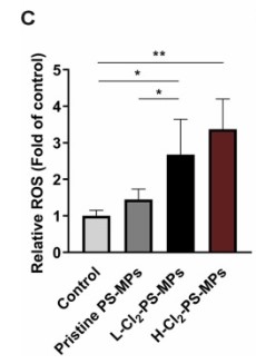
The last study of interest involved investigating the effect of chlorine on polystyrene microplastics. Since a good amount of PS-MPs can be removed from water through filtration, the impact of chlorine on PS-MPs remains unknown. However, it has been reported a significant portion of PS-MPs can pass through the filtration step of water treatment and onto a chlorine disinfection step, thus interacting with the PS-MPs. This study found that when PS-MPs was treated with chlorine, a significant increase in cell reactivity and toxicity was observed.
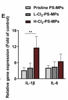
In the first figure (C), reactive oxygen species (ROS) is significantly increased in all chlorine-treated PS-MP groups compared to untreated (pristine) and control groups. In the second figure (E), PS-MPs treated with high amounts of chlorine (H-Cl2) resulted in significantly elevated IL-1beta, a gene that encodes the IL-1beta cytokine, suggesting that exposure to PS-MPs treated with H-Cl2 results in a change of in immune signaling.
IL-1beta: Cytokines are messengers of the immune system, and IL-1beta is a proinflammatory cytokine. Proinflammatory cytokines promote the secretion of inflammatory molecules and help prepare the immune cell to fight off foreign invaders.
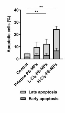
Another experiment involved looking at the percentage of apoptotic cells of different cell populations treated with the different PS-MPs or control. This figure portrays the results, and it was concluded that PS-MPs exposed to low or high chlorine levels significantly elevated the percentage of apoptotic cells suggesting that the exposure was cytotoxic. Future studies of this sort should be verified using pro-inflammatory markers like tumor necrosis factor-alpha (TNF-alpha).
The increasing abundance of microplastics is of the utmost concern, and future studies evaluating more effective and efficient methods of clearing microplastics from water are needed. As microplastics accumulate in the soil, they can be taken up into the roots of plants, and eaten, thereby passing the microplastics into the body. Microplastics can also be ingested via drinking water, contact with plastics, and even inhaled from the air. When the concentration of these microplastics increases, they have a negative impact on cell viability, DNA integrity, and inflammatory processes.
Common Microplastic Sources
Exposure to microplastics can vary drastically depending on location and lifestyle. A quite common source of microplastics, quite recently discovered is through inhalation. This source of microplastics is strongly correlated with location, with industrial cities having more airborne microplastics. Since leaving a city is not practical, here are a few more common sources of microplastics.
Although certain seafood dishes are more documented than others, mollusks are among the most microplastic-rich sea creature. If 300 grams of mollusk were consumed each week, an annual 89200 microplastic items would have been ingested from mollusks alone. It seems as if fish contain much less, however, with a lack of data supporting this claim, it should be taken with caution.
Another source of microplastics is salt. Several studies have found that sea salt contains microplastics derived from plastic waste found in the ocean. Salt is a major nutrient in the body and its intake is critical for regulating blood pressure, pH balance, and membrane ionization. Since salt cannot be avoided consumption of microplastics is inevitable, however, most people in the United States consume double the amount of salt recommended (10g vs 5g), thus doubling their exposure to microplastics.
The last major contributor of MPs is water, a non-negotiable liquid. The average American consumes 1.4 liters of water each day, with 23 microplastic items per liter, the annual ingestion equates to 11,753 microplastic items. This is water from water treatment plants, with tap water containing up to 61 microplastic items per liter, and bottled water with up to 1000 microplastic items per liter. With these upper limit values and an intake of 1.4 liters per day, tap water consumption results in 31,000 microplastic items per year, and bottled water consumption equates to 511,000 microplastic items per year. These are the upper limit values with the mean being roughly half of the reported value: 5,876 items/year for treated water, 15,500 items/year for tap water, and 255,500 items/year for bottled water consumers drinking 1.4 liters a day.
It should be noted that food contained within plastic wrap or packaging also contains microplastic items at around 11,000 microplastic items/year, with this number drastically increasing depending on the frequency of eating take-out or heating up food in packaging. Another important note is that the current method for detecting the presence of microplastic items is not optimal and future values will have to be adjusted in tandem with advances in detection technology.
Limiting Exposure to Microplastic Items
Hopefully, this article has not been too negative, as there are plenty of ways to limit exposure to microplastic items. The first being limit bottled water consumption, water in cans or from the tap already cuts the exposure to microplastics by more than 90% compared to individuals consuming all bottled water. The next step is to limit take-out and plastic storage. Since take-out containers and pre-packaged foods are packaged in plastic and oftentimes require heating, limiting consumption of these foods can cut microplastic exposure by 5,000 items per year. The last major lifestyle change is to limit salt intake, especially sea salt, to the recommended daily intake (5g), and in doing so can cut exposure by nearly 2,000 items per year. Exposure by inhalation is arguably the largest contributor to microplastic exposure, however, without better detection and data a proper method of limiting airborne microplastics is not well-supported.
Conclusion
Plastic is one of the world’s most commonly used packaging material and makes up 593 billion dollars in the United States alone. With the continued use of plastic, the advancements of recycling technology have not caught up, therefore leading to massive landfills and stretches of ocean engulfed by this malleable material. As wind, water, UV light, and mechanical forces slowly degrade this plastic, microplastic particles are released into the soil and drinking water. As temperatures rise, more plastic will be degraded and deposited into the body, therefore practical tools to limit the toxic effects of microplastic accumulation are needed. Some of these practices include limiting bottled water consumption, pre-packaged/processed foods, and regulating sea salt intake, however, global efforts must be made to improve recycling technology and limit microplastic deposition into drinking water, air, and the soil.
In America alone, 84 billion servings of tea were consumed in 2019, with 15% of that coming from green tea. Considering America is not the largest tea consumer, this is a staggering statistic that requires a cultural and biological explanation. Green tea, in particular, is advertised to relieve stress and anxiety yet energize the body significantly better than caffeinated drinks alone. A principal constituent of green tea is caffeine. However, another molecule, less well-known, is a major player in the stress relief associated with green tea. The compound is L-theanine or L-γ-glutamyl-ethyl amide, an amino acid analogue of L-glutamine (Gln, Q), found at around 25 mg/cup of green tea. L-theanine is considered safe in all mammalian cell lines and interestingly enough, the L form of theanine has special effects on neurological systems influencing anxiety. First, a brief introduction to what amino acids are and their responsibilities in the body. Secondly, a review of the literature highlighting L-theanine’s mechanism of action, and lastly, a brief discussion on the performance and cognitive benefits of L-theanine as seen through research.
Figure 1: L-theanine structure.
Figure 2: L-glutamine structure.
Amino Acids
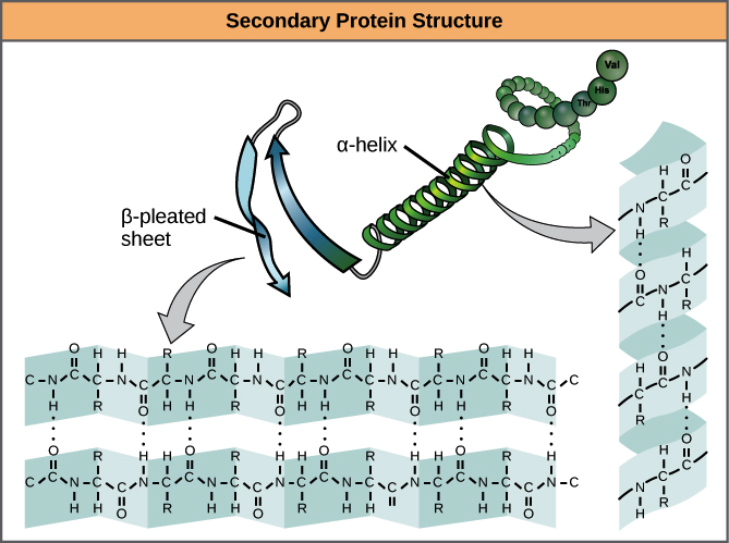
Amino acids are organic molecules containing a carboxyl and amino group. These small molecules are the building blocks of proteins. When two amino acids come together, a peptide bond is formed, thus forming a dipeptide. More amino acids can be added to this, and biologically this is completed from mRNA, tRNA, and ribosomes. Furthermore, peptides fold into alpha helices, beta-pleated sheets, and loops to form a protein’s secondary structure. These structures also have further interactions like hydrogen-hydrogen interactions, ionic bonds, and disulfide bonds courtesy of cysteine-cysteine bonding. However, even single amino acids are extremely important for cell signaling. For example, glutamate is an extremely important neurotransmitter, while leucine is a potent activator of mammalian target of rapamycin (mTOR). Altogether, amino acids can be building blocks for a larger protein, signaling molecules, and have other roles in nutrient sensing.
There are twenty essential amino acids, which are required to be taken in from the diet. However, not all amino acids are labeled as essential. Some amino acids like theanine are labeled non-proteinogenic amino acids, simply due to the fact that it is not used to build proteins but rather as signaling molecule. These kinds of amino acids can bind to or interact with receptors and other proteins to elicit a response or block a response from occurring.
Mechanism of Action
L-theanine is of special interest when it comes to productivity and stress relief particularly due to its impact on neurotransmission. For a drug to have any impact on psychological feelings or mood via the brain, it must be able to cross the blood-brain barrier and bind to specific receptors. An important characteristic of L-theanine administration is that brain levels start to rise, at the latest, 1-hour post-administration and brain levels don’t drop until 5-hours post-administration. This long time of action in the brain makes it a useful compound for administering prior to mental tasks.
L-theanine exerts its effect via antagonizing the glutamate receptor subtypes: AMPA receptor, kainate receptor, and NDMA receptor. Although studies report that L-theanine has a much lower affinity (80-fold less) than L-glutamic acid for the AMPA receptor, it seems it still exerts its effect through these glutamate receptor subtypes.
Glutamate is the primary excitatory neurotransmitter, thus by blocking the binding of glutamate to its receptors, L-theanine acts to decrease excitatory signaling. This occurs primarily in cortical neurons and this is the reason why L-theanine reduces anxiety. L-theanine has also been found to increase striatal dopamine and inhibit some aspects of norepinephrine signaling.
In this manner, L-theanine acts to block cyclic AMP (cAMP) formation which significantly decreases the impact norepinephrine has on stress and anxiety. L-theanine also seems to play an important role in altering the serotoninergic system, such that oral L-theanine increases serotonin concentrations in the hippocampus, striatum, and hypothalamus.
L-theanine is also quite commonly consumed prior to sleep due to its impact on gamma-aminobutyric acid (GABA) concentrations. Administration of 200-400 mg results in an increase in GABA concentrations thus resulting in higher-quality sleep. Lastly, the administration of L-theanine seems to be neuroprotective by preventing excess glutamate signaling or excitotoxicity. While some actions of L-theanine are still relatively unknown, it has many actions throughout the brain that impact several different signaling pathways. Based on the multifaceted mechanisms this compound is a promising supplement for boosting cognition and reducing stress and anxiety.
Benefits of L-theanine Supplementation
L-theanine is considered an anti-stress molecule, likely because it increases GABA, decreases excitatory glutamate neurotransmission, and reduces the impact of norepinephrine. A paper investigating psychological and physiological stress gave l-theanine to subjects and measured variation in heart rate. It was demonstrated that l-theanine reduced heart rate and variation of heart rate when compared to baseline and placebo measurements. This paper concluded that l-theanine is most likely decreasing sympathetic nervous system activity, or in other words, decreasing the impact of epinephrine and norepinephrine in the stress response. Another study using 20 healthy college students investigated the impact of l-theanine on subjective stress.
This study found that a dose of 200 mg/day of L-theanine was able to decrease subjective stress scores. The importance of this study resides in the fact that these subjects were labeled as healthy college adults, and a non-toxic, over-the-counter supplement was able to decrease stress is incredible. For further context, other supplements that claim to reduce
stress oftentimes only reduce stress in subjects prone to stress and anxiety. Another study used 34 healthy subjects, administered an L-theanine drink, and assessed stress levels over time. The study found that the l-theanine group (active) was significantly less stressed 1-hour post-consumption, but not 3-hours post-consumption.
There are also studies that use chronic restraint stress (CRS) to impair cognition in mice. This CRS-induced cognitive impairment results in abnormal corticosterone, the primary corticosteroid produced from the adrenal gland in rodents (cortisol in mammals), abnormal catecholamine levels in the brain, and other performance issues. A study performed in 2013 found that the administration of l-theanine alleviated this cognitive impairment.
In figure A, it was found that CRS mice not treated with l-theanine performed significantly worse in the morris water maze test. However, when treated with l-theanine at 2-4 mg/kg, the performance was not significantly different than normal healthy mice. Figure B demonstrated that mice treated with 4 mg/kg performed equally as well as normal healthy mice and significantly better than CRS mice and mice treated with l-theanine at 2 mg/kg. Overall, the main takeaway from these studies is that l-theanine treatment totally alleviated the cognitive impairment resulting from CRS.
L-theanine is a multifaceted compound, meaning its administration can potentially assist in a variety of diseases including Alzheimer’s disease (AD). A study published in 2018 investigated the impact of l-theanine on AD and the mechanism by which memory could be alleviated.
The first objective was to determine an action by which l-theanine may impact hippocampal memory systems, in which the authors found that treatment of l-theanine significantly raised phosphorylated PKA levels. To further confirm this, the researchers incubated the cells with SCH 23390, a D1 dopamine receptor antagonist, which confirmed that theanine acts through the D1 receptor to increase PKA phosphorylation.
The next set of experiments were aimed at discussing the impact of l-theanine on norepinephrine (NA) and dopamine levels in the hippocampus. The results indicate that l-theanine treatment raised both NA and dopamine levels in healthy mice and mice with AD. Altogether, the researchers concluded that l-theanine acts to promote hippocampal synaptic transmission and memory through the D1/PKA pathway, increase dopamine release, and increase NA release in the hippocampus.
A more recent 2022 study found that l-theanine was able to alter the gut microbiota, thus impacting lipid metabolism in a positive manner. It was found that through l-theanine administration, Lactobacillus, Streptococcus, Bacteroides, Clostridium, and Enterorhabdus were decreased and serum triglycerides and cholesterol were significantly decreased as a result.
The results indicated above suggest that l-theanine at 100-300 mg/kg significantly reduces serum total cholesterol (A), triglycerides (B), LDL-cholesterol (C), liver total cholesterol (E), and liver triglycerides (F). It was also found that serum and liver HDL-cholesterol were significantly increased compared to the control. These results indicate that l-theanine has a positive influence on lipid profiles in mice and this warrants future studies.
While l-theanine has antioxidant properties, metabolic regulatory properties, and other benefits, it is mostly taken in conjunction with caffeine to improve performance. While an earlier discussion focused on memory improvement after administration of l-theanine, its stress-reducing properties are especially beneficial when taken with caffeine. Since caffeine oftentimes results in jitters and anxiety, co-administration with l-theanine reduces jitters and caffeine-induced anxiety.
Experiments from a paper found that the theanine + caffeine group performed significantly better in the “new word” experiment. Along with this, the theanine + caffeine group also had significantly reduced response time compared to placebo and caffeine-only groups. Another paper found that subjects given l-theanine plus caffeine had reduced jitters and increased alertness compared to theanine only, caffeine alone, and placebo groups. Another double-blind, placebo-controlled study found that caffeine plus theanine significantly improved attention tasks and overall mood ratings suggesting that this combination is effective for enhancing work productivity.
Theanine for Enhancing Productivity
The findings from the literature as listed above suggest that l-theanine is a useful compound when combined with caffeine. The combination of these compounds provides calm alertness, with the alertness coming from the caffeine, and the calm coming from the l-theanine. A cup of green tea contains around 25 mg, which is a suboptimal amount of l-theanine. Other studies use 50 mg, but the most common dose used is anywhere from 100-500 mg, with doses upwards of grams being taken without any side effects. A dose of 100-200 mg caffeine paired with 200 mg l-theanine is a useful combination when enhancing work productivity.
Another useful compound to pair with l-theanine is theacrine. As described in a previous article (Theacrine Review), theacrine is similar to caffeine, except with a more pronounced impact on neurotransmission and a longer half-life. Since caffeine and theacrine both primarily act on the adenosine receptor, they both increase alertness, thus any stress or anxiety coming from theacrine will be alleviated via l-theanine.
Of course, l-theanine can be used alone to decrease stress. For days not focused on long work hours, l-theanine can be administered alone to decrease stress and allow for caffeine desensitization.
Vitamin D has gained enormous notoriety over the past decade, primarily due to correlation studies finding that a large portion of the population is vitamin D-deficient. Since then, study after study have found correlations between vitamin D and disease states such as heart disease and Alzheimer’s. Vitamin D is a steroid-based molecule, and its classification as a vitamin occurred in 1920, due to the notion that a lack of vitamin D can cause rickets, thus its intake is vital for health. However, more recently, it was discovered that vitamin D is multifaceted, that is, vitamin D has actions on calcium and bone homeostasis, sex hormone production, and inflammation processes. The structure of vitamin D is more similar to pregnenolone, a cholesterol derivative, than to vitamin K and other vitamins. Since vitamin D is synthesized from cholesterol, it is a fat-soluble molecule able to cross the cell membrane and bind to nuclear receptors that act to influence gene transcription.
Vitamin D can be taken in from the diet; it is usually found in fish; it can be endogenously produced when the skin is exposed to ultraviolet light. After the conversion from UV light, enzymes produced and released from the liver and kidneys can convert the inactive form to the active vitamin D3 or calcitriol. In the active form, calcitriol can bind to the vitamin D receptor (VDR), which may interact with other receptors such as the retinoid X receptor (RXR), which can control the transcription of certain genes. Vitamin D plays a major role in calcium-phosphorus homeostasis at the absorption and transcriptional level, making it a multifaceted molecule.
There have been studies conducted throughout the decade that explore the actuality of previously conducted correlation studies. The model “one-size fits all” is a statement that holds true, not only in certain regions of science but in all matters of exploration. With the advancement of population and genetic studies, it is becoming increasingly known that circulating levels of vitamin D are different in different groups of people. From a logical perspective, the ancestors living in the northern regions were exposed to less UV light than ancestors living near the equator. In addition, clothing needed to keep warm oftentimes renders this UV light useless in the winter months. Also, dietary sources of vitamin D are not as abundant as previously thought, and for a hunter-gatherer group, it would be logical to think these ancestors were not receiving what we deem today a recommended daily intake of vitamin D. Besides the fact that fat-soluble vitamins can be stored in adipose tissue for long-term use, including vitamin D, genetics, and epigenetics point to another solution as to why vitamin D deficiency did not severely impact our ancestors. Genetics and epigenetics allow receptors to elicit a response intensity, such that low levels of vitamin D in a northern ancestor could facilitate an adequate response intensity, similar to that of high vitamin D in an ancestor closer to the equator. The notion that a reference range is adequate is nonsensical and has led to people taking vitamin D supplements that may contain much higher levels of vitamin D than needed, possibly harming their health.
With the necessary background, this journal review aims to investigate a recent article focusing on the negative impact of vitamin D supplementation on Alzheimer’s disease. Alzheimer’s disease (AD) is a rather complex disease, and even more so after recent scandals involving the pathology of the disease. The pathology of AD is rather extensive, but briefly, it is a neurodegenerative disease in which amyloid beta and tau tangles begin to dominate, cause inflammation, and eventually lead to neuronal death. This cell death, or infiltration of foreign proteins, causes signal disruption, which is clinically referred to as memory loss. The mechanisms behind Alzheimer’s disease are extremely complex and recent findings indicate that vitamin D levels may be decreased because of Alzheimer’s onset instead of what was previously described, which was that vitamin D deficiency causes Alzheimer’s.
This study is very comprehensive encompassing multiple animal models and a large population study investigating VD and AD. The first model acted to investigate whether VD deficiency is caused by AD or if, as correlation studies concluded, VD deficiency causes AD. To determine this, two groups were established, a control group and an APP/PS1 Alzheimer’s group (TG). The researchers took serum vitamin D levels at 2, 4, and 6 months old to determine at which point VD. The results of this experiment showed that as AD progresses, serum VD decreases, suggesting that AD causes VD deficiency. Immunofluorescence showed that there are significantly more amyloid beta plaques in Alzheimer’s mice given a vitamin D supplement when compared to mice fed a normal diet. This trend also follows with Glial fibrillary acidic protein (GFAP), a marker for AD progression. Next, the researchers used a western blot, which is a technique used to determine if a protein is expressed. The western blot was looking at amyloid beta protein, beta secretase-1 (BACE), and a control protein GAPDH. The results indicate that in the hippocampus of Alzheimer’s-induced mice, a diet supplemented with VD resulted in higher amyloid beta plaques and BACE protein levels. To test if VD supplementation led to worse cognitive impairment in AD, a Morris water maze test was conducted and the results demonstrate that VD supplementation worsened the time of escape.
Figure 1: Serum vitamin D3 in APP/PS1 (Alzheimer’s) (TG) and wild-type (WT) mice at 2, 4, and 6 months.
Figure 2: Western blot of VDR, apoptotic and autophagic marker proteins in SH-SY5Y cells exposed to Aβ42 or plus without or without Vitamin D3.
Figure 3: Immunofluorescence of amyloid beta plaques or GFAP in control cells or vitamin D-supplemented cells.
After these results, the authors then concluded that indeed the animal models pointed to the fact that AD causes VD deficiency and that supplementing with VD amidst Alzheimer’s seems to worsen the marker proteins and behavior. The next step of this study was to investigate by which mechanism is AD using or causing VD deficiency and how increasing VD via supplementation worsens AD progression. Under normal physiological conditions, when VD binds to the VDR, it binds to the retinoid X receptor, which then activates transcription via promoting transcription factor colocalization. However, existing research conducted by this group demonstrated that AD conditions may cause the VDR when bound by VD to form a complex with p53. This p53-VDR complex seems to promote pathological conditions within the cell.
To determine if the hypothesis is correct the authors decided to test whether VD supplementation improved VDR-RXR complex formation in physiological or pathophysiological conditions. The results from this experiment showed that VD supplementation in AD conditions does not rescue VDR-RXR formation. However, in normal physiological conditions, VD supplementation increased VDR-RXR complex levels two-fold compared to physiological conditions given no VD supplement. The next two western blots show that in hippocampal cells of AD mice given VD supplementation, a p53-VDR complex becomes predominant. The next few graphs demonstrated that AD complications are increased due to the formation of a VDR-p53 complex because when cells are treated with a PTF-alpha, a p53 inhibitor, amyloid beta plaques and GFAP-positive cells are totally alleviated when compared to cells not treated with the inhibitor.
Figure 4: (a) Mammalian two-hybrid assays for studies of the interaction of VDR with RXR in neurons exposed to amyloid beta plus with vitamin D3. (b) Western blot demonstrating the presence of amyloid beta plaques and vitamin D supplementation increases the abundance of p53. (c) Western blot showing mice with an Alzheimer’s disease phenotype have increased p53 expression when supplemented with vitamin D.
So far, the paper has concluded some major points about AD that have been quite controversial. Firstly, the data from this paper hints at the fact that VD supplementation increases GFAP-positive cells and amyloid beta plaques when supplementation is started during the onset of AD. Secondly, it also seems like VD supplementation during the onset of AD increases the formation of VDR-p53 complexes which play a role in AD pathophysiology.
To elevate the claims the authors made, a large population human study using 14,648 subjects over the age of 65 and dementia-free. The findings from the large-population study demonstrate that a low dose of VD did not impact the incidence of dementia. However, a large dose or medium dose of VD significantly elevated the risk of dementia. A similar study using 980 patients with preexisting dementia were followed for 11 years and compared the effects of a VD supplementation on mortality. It was found that a high dose of VD increased the mortality of dementia by 2-fold. However, it was found that a medium or low dose did not impact mortality. The findings of the large-population study need further validation, better controls, and a clinical trial to be conclusive. The findings could be indicative of the issues with a VD supplement in patients with preexisting dementia or patients prone to dementia. In conclusion, a vitamin D supplement in the elderly seems to have beneficial impacts on bone health but may negatively affect neurological health. The in vitro and in vivo experiments in this study are promising and require more validation because several other papers have found conflicting results yet are not as comprehensive as this research.
Figure 5: Classical view of vitamin D on neuronal cells in the brain.
Hello everyone,
My name is Joshua Giblin. I am a post-bachelor researcher/research technician at USC. My interests range from nutrition to nanomedicine and also practical science to improve everyday life. Through this blog, I aim to communicate practical scientific research and present it to curious individuals so that an educated decision can be made. Thank you for reading the blog and showing your support.
Literature cited
Chatterjee, P., Pedrini, S., Stoops, E., Goozee, K., Villemagne, V. L., Asih, P. R., Verberk, I. M. W., Dave, P., Taddei, K., Sohrabi, H. R., Zetterberg, H., Blennow, K., Teunissen, C. E., Vanderstichele, H. M., & Martins, R. N. (2021). Plasma glial fibrillary acidic protein is elevated in cognitively normal older adults at risk of Alzheimer’s disease. Translational Psychiatry, 11(1), Article 1. https://doi.org/10.1038/s41398-020-01137-1
Lai, R.-H., Hsu, C.-C., Yu, B.-H., Lo, Y.-R., Hsu, Y.-Y., Chen, M.-H., & Juang, J.-L. (2022). Vitamin D supplementation worsens Alzheimer’s progression: Animal model and human cohort studies. Aging Cell, 21(8), e13670. https://doi.org/10.1111/acel.13670
Nishikawa, M., Yasuda, K., Takamatsu, M., Abe, K., Okamoto, K., Horibe, K., Mano, H., Nakagawa, K., Tsugawa, N., Hirota, Y., Horie, T., Hinoi, E., Okano, T., Ikushiro, S., & Sakaki, T. (2020). Generation of novel genetically modified rats to reveal the molecular mechanisms of vitamin D actions. Scientific Reports, 10(1), Article 1. https://doi.org/10.1038/s41598-020-62048-1